35 gram positive bacteria diagram
Bacilleus cereus. food poisoning, reheated rice syndrome, N/V within 1-5 hr, watery, nonbloody diarrhea + GI pain (8-18 hr) Listeria monocytogenes. the only G+ to produce endotoxin;amnionitis, septicemia, spontaneous abortion in preg & on steroid; พบใน frozen food & UHT products. long, branching filaments. the difference is clear but in simple explanation gram staining is what makes bacteria to be gram positive or negative and this happens because gram positive bacteria have thick peptidoglycan which retains crystal violet staining dye as opposed to gram. Reply. Mowaddah khan.
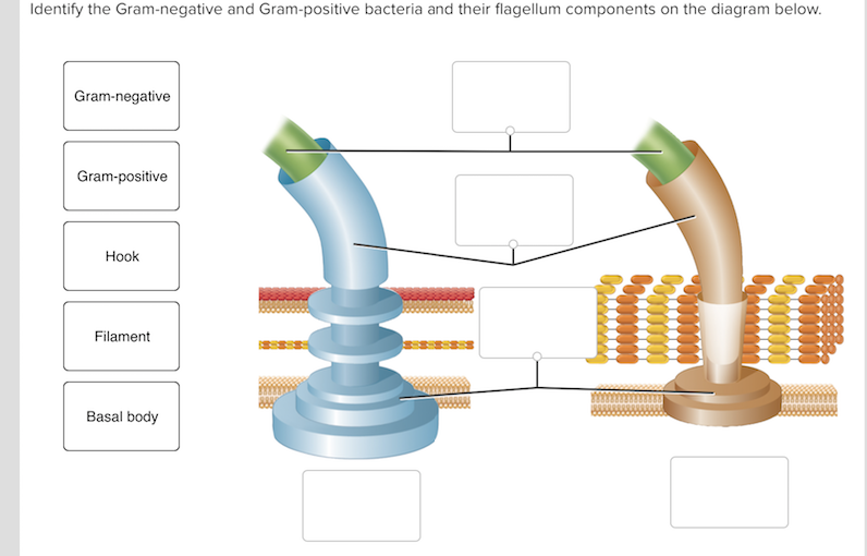
S. pyogenes is a gram-positive bacterium. Gram-positive bacteria have structural differences that distinguish them from gram-negative organisms. Of particular interest to this case is the fact that these differences in structure can be exploited during treatment. Drag each of the labels onto the diagram to correctly label the indicated structures.
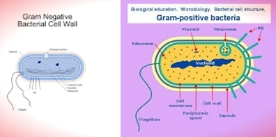
Gram positive bacteria diagram
For the gram-positive cell wall, it has a thickness of about 20-80nm thickness made up of a thick peptidoglycan layer outside its cell membrane, unlike the thin layer of gram-negative bacteria (10-15nm) which has a very thin layer of the peptidoglycan of 2-7nm but has a thicker lipid layer making it quite complex than the Gram-positive cell wall. Both Gram-positive and Gram-negative bacteria take up the same amounts of crystal violet (CV) and iodine (I). The CV-I complex, however, is trapped inside the Gram-positive cell by the dehydration and reduced porosity of the thick cell wall as a result of the differential washing step with 95 percent ethanol or other solvent mixture. In Gram-positive bacteria, peptidoglycan makes up as much as 90% of the thick cell wall enclosing the plasma membrane. Gram Staining! During Gram staining, these thick, multiple layers (20-80 nm) of peptidoglycan retain the dark purple primary stain crystal violet, whereas Gram-negative bacteria stain pink.
Gram positive bacteria diagram. Gram-positive Cell Wall. Gram-negative Cell Wall. Outer Membrane. Cytoplasmic Membrane. Membrane Proteins. Porin Flow Chart Of The Study Design Each Patient Having A First Positive Scientific Diagram. Flowchart For Identification Of Anaerobic Gram Positive Bacilli 1 Scientific Diagram. Gram Negative Bacteria Positive Microbiology Flowchart Others Angle Text Infection Png Pngwing. Starting With Figure 17 5 And Using From Case Study T Chegg. Pathogenic bacteria are bacteria that can cause disease. This article focuses on the bacteria that are pathogenic to humans. Most species of bacteria are harmless and are often beneficial but others can cause infectious diseases.The number of these pathogenic species in humans is estimated to be fewer than a hundred. By contrast, several thousand species are part of the gut flora present in ... Gram-positive bacteria are surrounded by many layers of peptidoglycan (PG), which form a protective shell that is 30–100 nm thick (Silhavy et al. 2010). The PG ...
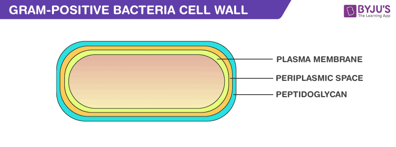
Gram positive bacteria are a group of organisms that fall under the phylum Firmicutes (however, a few species have a Gram negative cell wall structure). As compared to Gram negative bacteria, this group of bacteria is characterized by their ability to retain the primary stain (Crystal violet) during Gram staining (giving a positive result). ... Download scientific diagram | Antibiotics used for gram positive and gram negative bacteria with its disc content from publication: Antibiotic sensitivity profile of bacterial pathogens in ... The Gram-Positive Cell Wall. As mentioned in the previous section on peptidoglycan, Gram-positive bacteria are those that retain the initial dye crystal violet during the Gram stain procedure and appear purple when observed through the microscope. As we will learn in lab, this is a result of the structure and function of the Gram-positive cell wall. Compare and contrast the cell walls of typical Gram-positive and Gram-negative bacteria. 3. Relate bacterial cell wall structure to the Gram-staining reaction. 37 . 38 Bacterial Cell Wall • Peptidoglycan (murein) -rigid structure that lies just outside the cell plasma membrane
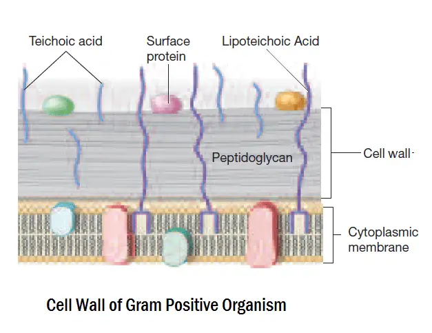
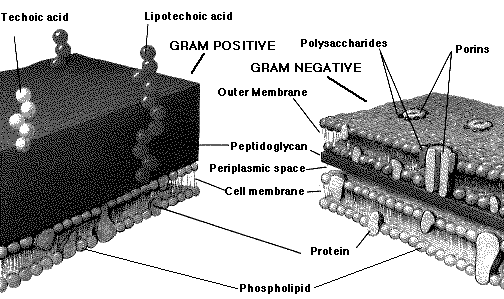
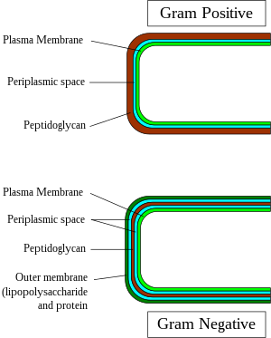
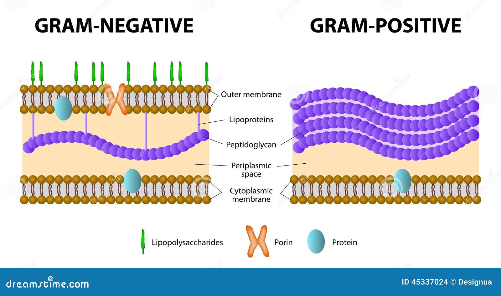
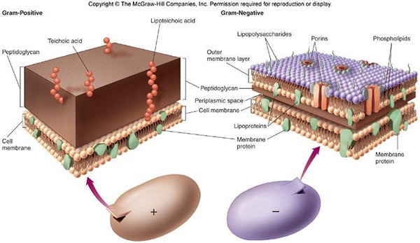
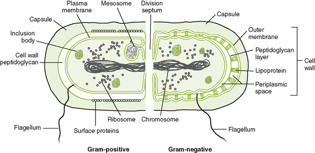
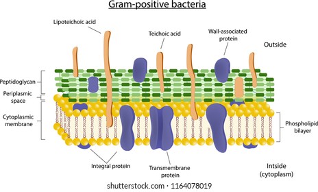
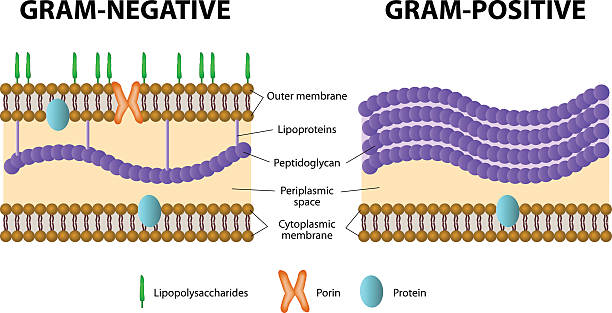
In the course of evolution, Gram-positive bacteria, defined here as prokaryotes from ... bacteria: from single protein to macromolecular protein structure. The diagram below illustrates the differences in the structure of gram positive and gram negative bacteria. The difference is clear but in simple explanation gram staining is what makes bacteria to be gram positive or negative and this happens because gram positive diagram. The diagram below illustrates the differences in the structure of gram ... THE GRAM POSITIVE CELL WALL. The Gram positive cell wall has a thick peptidoglycan (orange red in this picture) layer outside the plasma membrane.There may be a gap or periplasmic space between the peptidoglycan layer and the plasma membrane.Various membrane proteins can be seen floating in the plasma membrane.Elongate molecules called teichoic acids intermesh with the peptidoglycan layer. Gram-positive bacteria remain dark-violet or purple coloured, but Gram-negative bacteria become red (pink) (Fig. 2.10). This grouping does not depend on the shape of bacteria, e.g., the bacillus-shaped bacteria like Bacillus subtilis is Gram-positive, while the other rod-shaped bacteria like Coxiella burnetii is Gram-negative.
Gram-positive bacteria are classified by the color they turn after a chemical called Gram stain is applied to them. Gram-positive bacteria stain blue when ...
The cell wall structure of Gram negative bacteria is more complex than that of Gram positive bacteria. Located between the plasma membrane and the thin peptidoglycan layer is a gel-like matrix called periplasmic space. Unlike in Gram positive bacteria, Gram negative bacteria have an outer membrane layer that is external to the peptidoglycan ...
The gram-Positive Cell wall of Bacteria Bacterial cell wall that is gram-positive contains peptidoglycan and teichoic acids with some species having additional carbohydrates and proteins. The murein component is what gives shape to the gram-positive bacterial cell wall; it also helps the bacteria cells to resist osmotic pressure.

Protein Secretion And Surface Display In Gram Positive Bacteria Philosophical Transactions Of The Royal Society B Biological Sciences
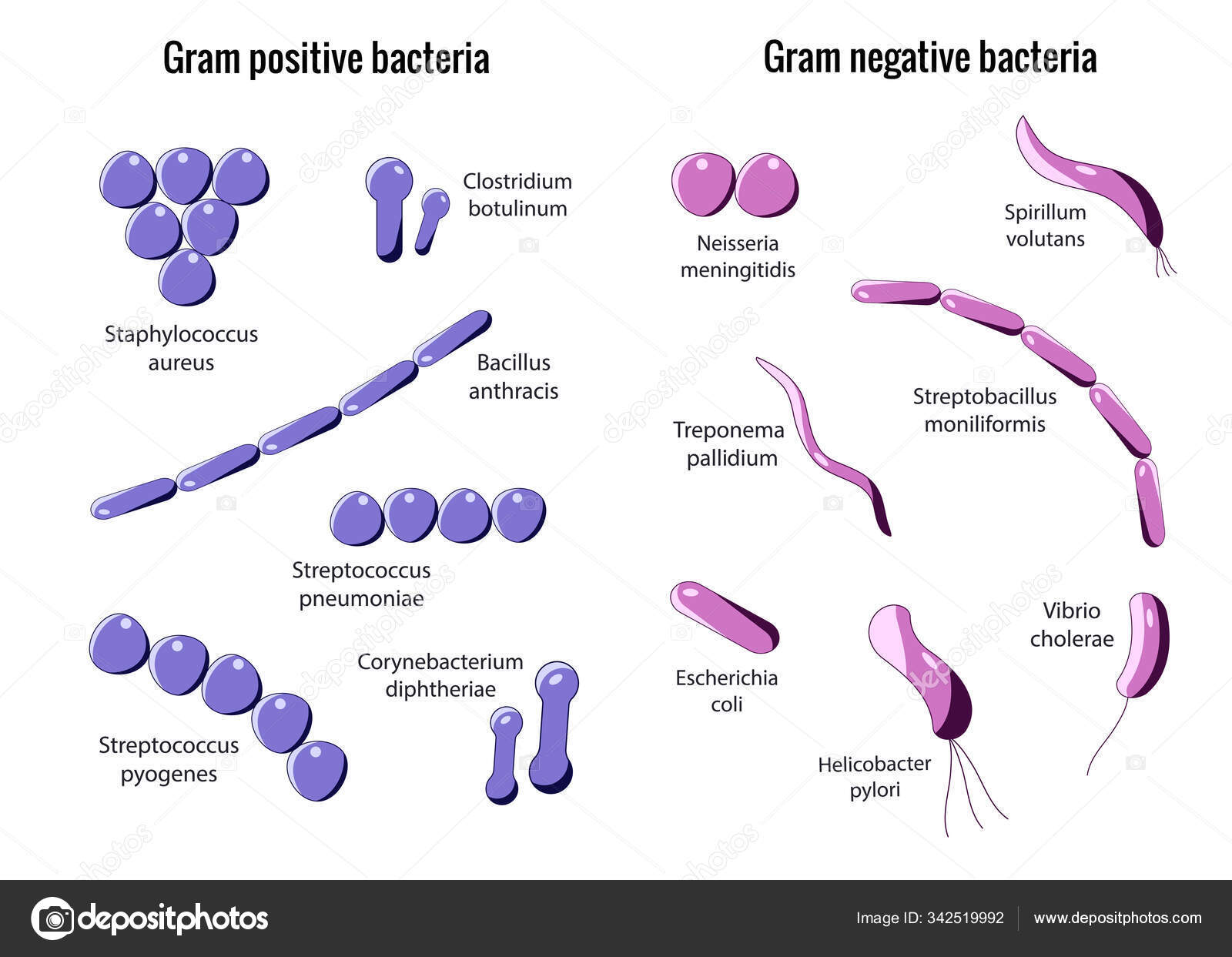
Gram-positive bacteria comprise cocci, bacilli, or branching filaments. Health professionals need to understand the important difference between gram-positive and gram-negative bacteria. Gram-positive bacteria are bacteria classified by the color they turn in the staining method. Hans Christian Gram developed the staining method in 1884.
Though cell wall and chemical structure are different in gram-positive and gram-negative bacteria. In gram-positive bacteria, the cell wall is thick and homogeneous but the cell wall of gram-negative bacteria is relatively lesser thick and heterogeneous. *** Functions: The cell wall is responsible for the shape of the bacterial cells.
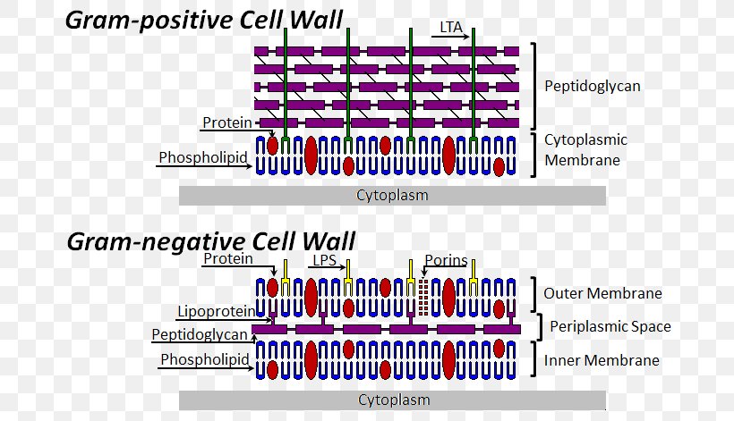
Bacterial Cell Structure Cell Wall Gram Positive Bacteria Gram Negative Bacteria Png 696x471px Bacterial Cell Structure
4 Bacteria: Cell Walls . It is important to note that not all bacteria have a cell wall.Having said that though, it is also important to note that most bacteria (about 90%) have a cell wall and they typically have one of two types: a gram positive cell wall or a gram negative cell wall.. The two different cell wall types can be identified in the lab by a differential stain known as the Gram stain.
Gram-positive bacteria contain many surface proteins (e.g. protein A, fibrinonectin-binding ...
Gram positive bacteria that have been overly heat fixed resulting in destruction of all or parts of their cell wall can appear to be pink (Gram negative) or have pink areas. This is an artifact! Further, old moribund cultures of Gram positive cells can appear pink. This is because the cell wall has allowed the challenge rinse to enter the cell. ...
In bacteriology, gram-positive bacteria are bacteria that give a positive result in the Gram stain test, which is traditionally used to quickly classify bacteria into two broad categories according to their type of cell wall.. Gram-positive bacteria take up the crystal violet stain used in the test, and then appear to be purple-coloured when seen through an optical microscope.
Gram-positive bacteria are bacteria with thick cell walls. In a Gram stain test, these organisms yield a positive result.
Gram positive bacteria have a thick cell wall, which consists of up to around 30 layers of peptidoglycan. This cell wall surrounds a ...
Gram-positive bacteria are the genus of bacteria family and a member of the phylum Firmicutes. These bacteria retain the colour of the crystal violet stain which is used during gram staining. These bacteria give a positive result in the Gram stain test by appearing purple coloured when examined under a microscope, hence named, gram-positive ...
Mycoplasma are bacteria that have no cell wall and therefore have no definite shape. Outer Membrane: This lipid bilayer is found in Gram negative bacteria and is the location of lipopolysaccharide (LPS) in these bacteria. Gram positive bacteria lack this layer. LPS can be toxic to a host and can stimulate the host's immune system.
The cell wall of Gram-positive bacteria is a complex assemblage of glycopolymers and proteins. It consists of a thick peptidoglycan sacculus ...
Christian Gram, a Danish Physician in 1884 developed a staining technique to distinguish two types of bacteria. The two categories of bacteria based on gram staining are Gram positive bacteria and Gram negative bacteria. Bacteria are first stained with crystal violet or gentian violet.
8. According to Peberdy (1980) the only compound present in the cell walls of both Gram-negative and Gram-positive bacteria is 'peptidoglycan'. The cell walls of Gram-positive bacteria contain up to 95% peptidoglycan and up to 10% teichoic acids. 9. Cytoplasmic membrane is a thin (5-10 nm) layer lining the inner surface of the cell wall.
As Gram positive bacteria lack an outer lipid membrane, when correctly referring to their structure rather than staining properties, are termed ...
Gram positive bacteria have a large peptidoglycan structure. As noted above, this accounts for the differential staining with Gram stain. Some Gram positive bacteria are also capable of forming spores under stressful environmental conditions such as when there is limited availability of carbon and nitrogen. Spores therefore allow bacteria to ...
Gram-positive bacteria, on the other hand, retains the gram stain and show a visible violet colour upon the application of mordant (iodine) and ethanol (alcohol). Gram-positive bacteria constitute a cell wall, which is mainly composed of multiple layers of peptidoglycan that forms a rigid and thick structure.
In Gram-positive bacteria, peptidoglycan makes up as much as 90% of the thick cell wall enclosing the plasma membrane. Gram Staining! During Gram staining, these thick, multiple layers (20-80 nm) of peptidoglycan retain the dark purple primary stain crystal violet, whereas Gram-negative bacteria stain pink.
Both Gram-positive and Gram-negative bacteria take up the same amounts of crystal violet (CV) and iodine (I). The CV-I complex, however, is trapped inside the Gram-positive cell by the dehydration and reduced porosity of the thick cell wall as a result of the differential washing step with 95 percent ethanol or other solvent mixture.
For the gram-positive cell wall, it has a thickness of about 20-80nm thickness made up of a thick peptidoglycan layer outside its cell membrane, unlike the thin layer of gram-negative bacteria (10-15nm) which has a very thin layer of the peptidoglycan of 2-7nm but has a thicker lipid layer making it quite complex than the Gram-positive cell wall.
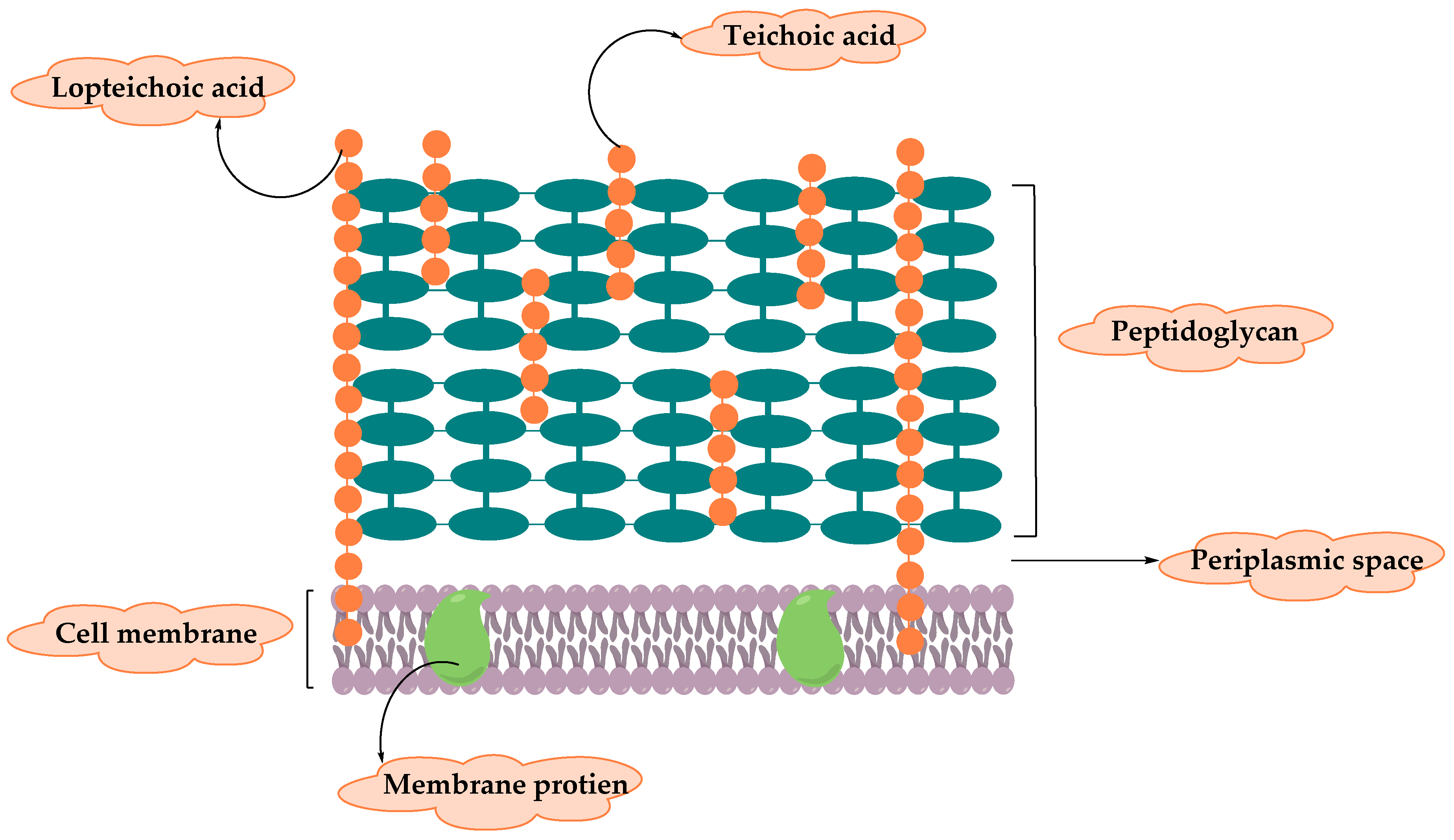
Molecules Free Full Text Resistance Of Gram Positive Bacteria To Current Antibacterial Agents And Overcoming Approaches
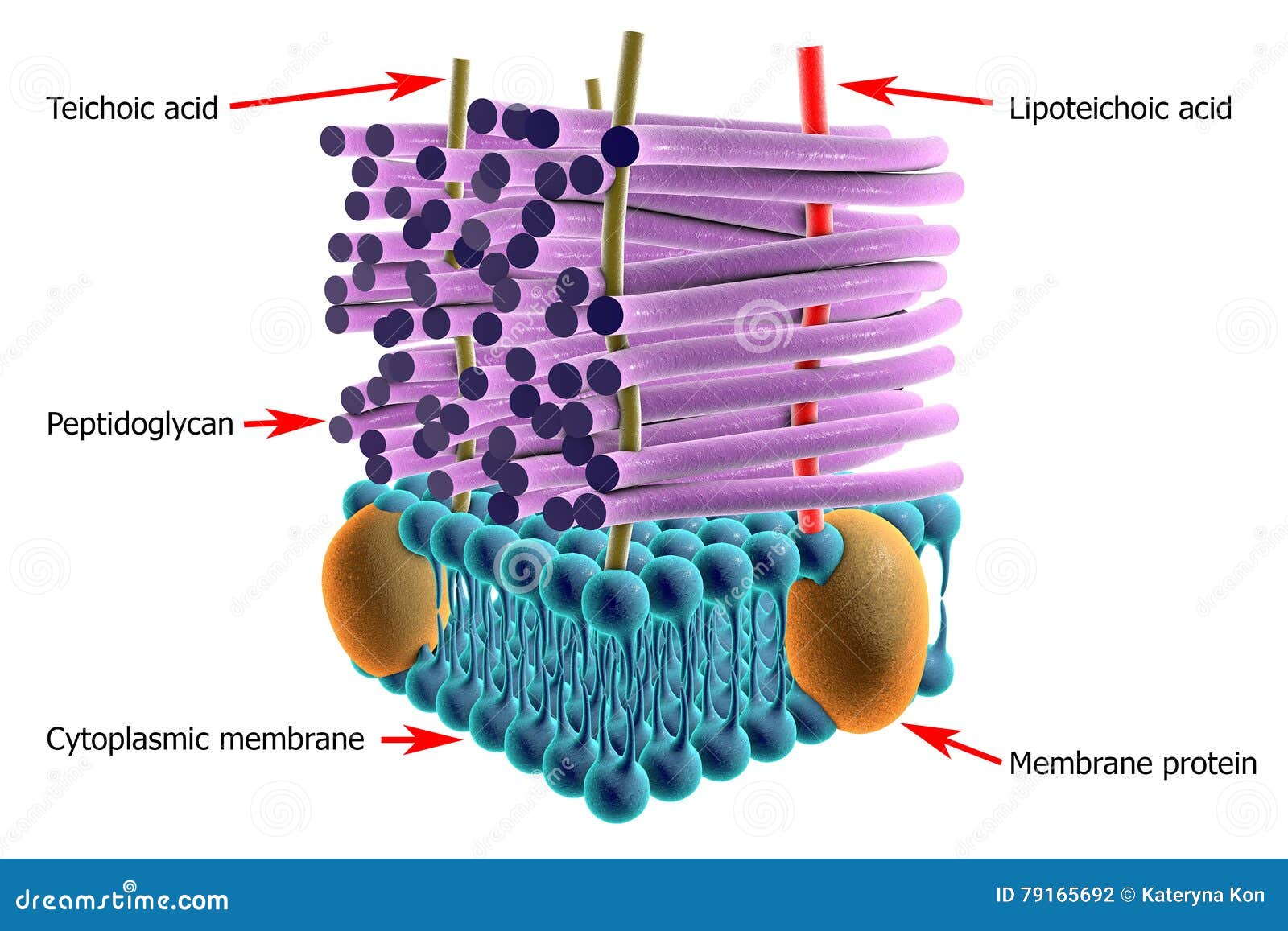
Structure Of Gram Positive Bacteria Cell Wall Stock Illustration Illustration Of Microbial Muramic 79165692




:max_bytes(150000):strip_icc()/gram_positive_bacteria-5b7f3032c9e77c0050f88457.jpg)







0 Response to "35 gram positive bacteria diagram"
Post a Comment