38 simple columnar epithelium labeled diagram
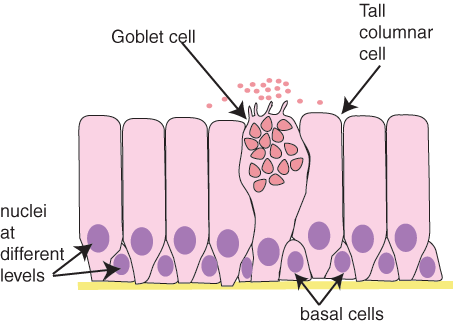
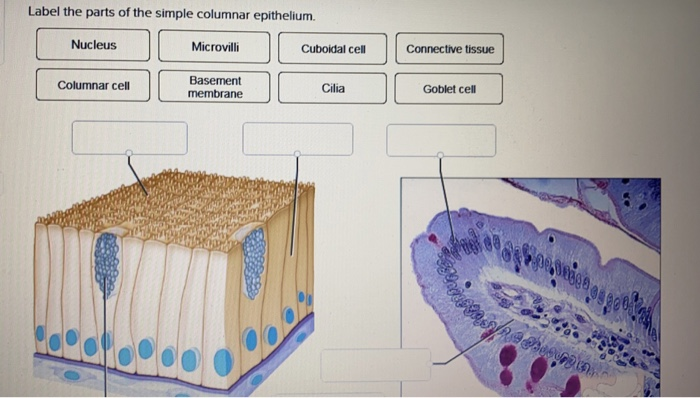
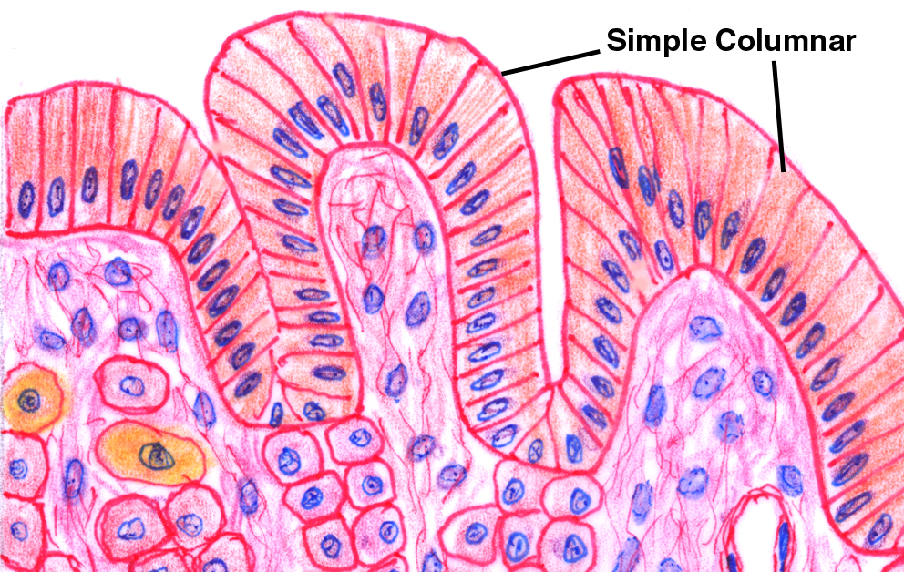
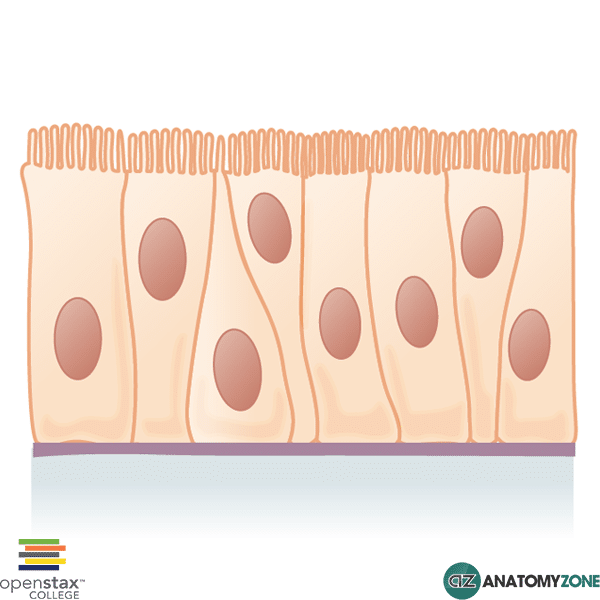
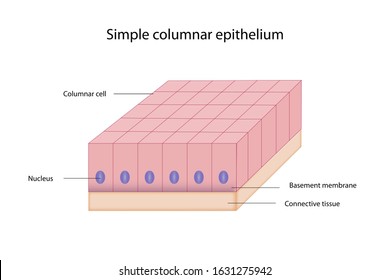
Simple Columnar Epithelium Definition. Simple columnar epithelia are tissues made of a single layer of long epithelial cells that are often seen in regions where absorption and secretion are important features. The cells of this epithelium are arranged in a neat row with the nuclei at the same level, near the basal end. Columnar epithelia, which form the lining of the digestive tract, can be either simple or stratified. The cells are long and narrow. The nucleus is elongated and located on the basal side of the cell. Ciliated columnar epithelium is composed of simple columnar epithelial cells that display cilia on their apical surfaces.
A simple columnar epithelium is a columnar epithelium that is uni-layered. In humans, a simple columnar epithelium lines most organs of the digestive tract including the stomach, . Simple Columnar Epithelium: A Labeled Diagram and Functions Epithelium is a tissue that lines the internal surface of the body, as well as the internal organs.

Simple columnar epithelium labeled diagram
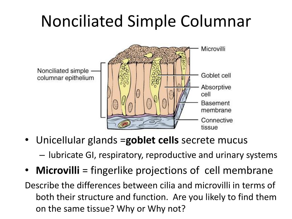
The simple columnar epithelium is either ciliated or non-ciliated, and the non-ciliated columnar epithelium has microvilli on the apical area. Simple Columnar Epithelium Structure. The simple columnar epithelium is composed of a single layer of elongated cells that are always taller than they are wide and are linked directly to the basement ... Simple Epithelium- it is composed of one layer of a cell and mostly has a secretory or an absorptive function. Compound (Stratified) Epithelium- it is made up of two or more than two layers of cells and mostly has a protective function. The glandular epithelium is made up of cuboidal or columnar cells. They are specialised for secretion. Start studying Simple Columnar Epithelium. Learn vocabulary, terms, and more with flashcards, games, and other study tools.
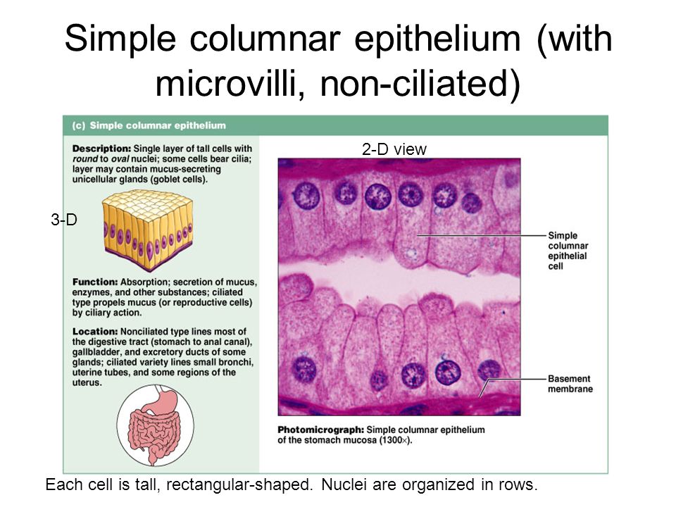
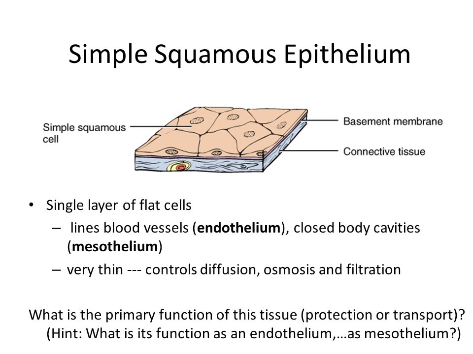
Simple columnar epithelium labeled diagram. At the junction with the esophagus, the stratified squamous epithelium of the esophagus abruptly changes to the simple columnar epithelium of the stomach. Simple columnar epithelium is recognizable by the shape and position of the nuclei. They are elongated and arranged in a neat, single row along the basement membrane. Figure 6: Simple ... Simple cuboidal epithelium is found in glandular tissue and in the kidney tubules. Simple columnar epithelium lines the stomach and intestines. Pseudostratified columnar epithelium lines portions of the respiratory tract and some of the tubes of the male reproductive tract. Transitional epithelium can be distended or stretched. Simple columnar epithelium. Simple columnar epithelium consist of a single layer of cells that are taller than they are wide. This type of epithelia lines the small intestine where it absorbs nutrients from the lumen of the intestine. Simple columnar epithelia are also located in the stomach where it secretes acid, digestive enzymes and mucous. The simple squamous epithelium is different from other types of epithelial tissue such as simple cuboidal, simple columnar, and stratified squamous epithelium in that it is only made of one layer ...
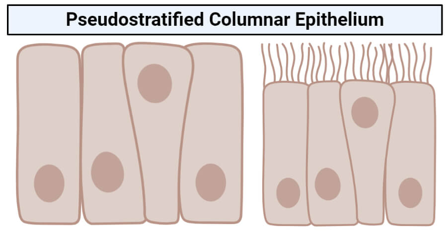
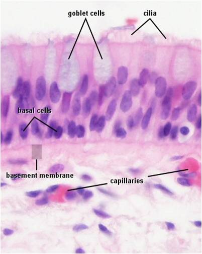
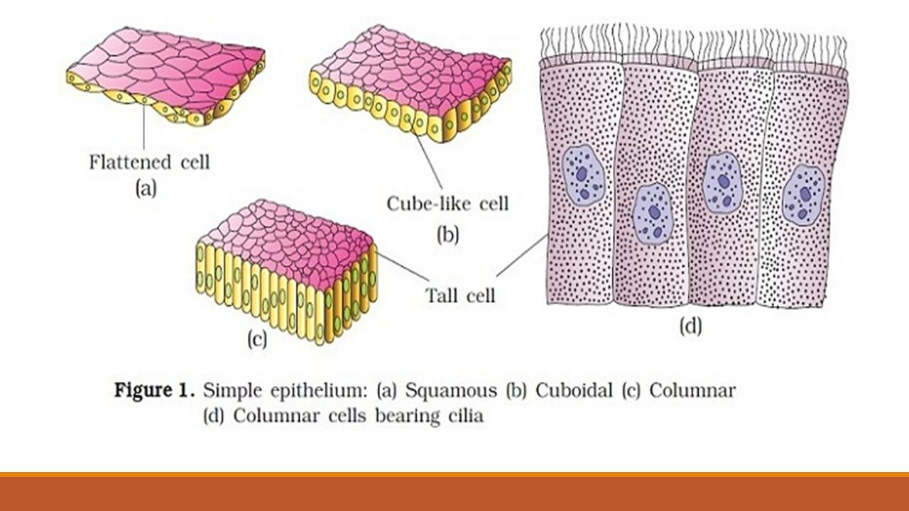
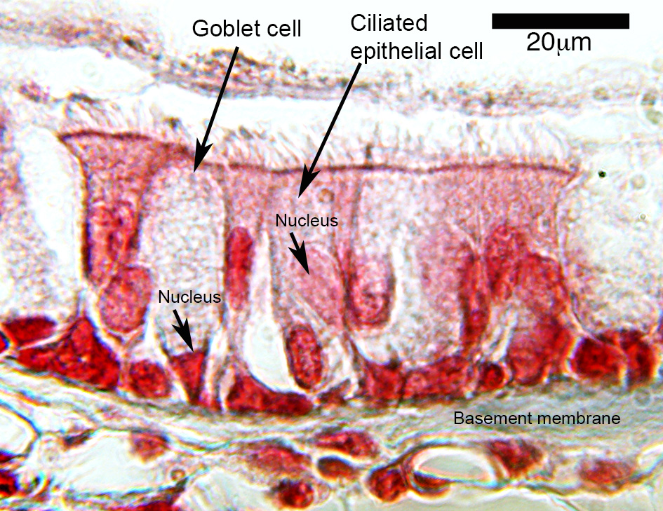
Simple epithelium can be divided into 4 major classes, depending on the shapes of constituent cells. The cells found in this epithelium type are flat and thin, making simple squamous epithelium ideal for lining areas where passive diffusion of gases occur.Areas where it can be found include: skin, capillary walls, glomeruli, pericardial lining, pleural lining, peritoneal cavity lining, and ... Simple Cuboidal Epithelium (Figure 4.3b) Simple cuboidal epithelium consists of a single layer of cube-shaped cells. This epithelium forms the secretory cells of many glands, the walls of the smallest ducts of glands, and the walls of many tubules in the kidney. Its functions are the same as those of simple columnar epithelium. These labelled diagrams should closely follow the current Science courses in histology, anatomy and embryology and complement the virtual microscopy used in the current Medical course. ... and simple columnar epithelium with basal striations FEATURE: lobe, lobules and ducts (TS) Simple secretory columnar epithelium lines the stomach and uterine cervix.The simple columnar epithelium that lines the intestine also contains a few goblet cells. In histological slides of pseudostratified epithelium , it looks as though some of the cells are not in contact with the basal lamina, and the nuclei are at different levels.
Simple Columnar Epithelium Labeled Diagram and Function Microscope Simple Squamous Epithelium Labeled Diagram Written By MacPride Friday, December 25, 2020 Add Comment Edit. ... Vintage Microscope Slide Mammal Simple Columnar Epithelium 31 2420. Epithelial Tissue Springerlink. Simple Columnar Epithelium. Simple Epithelium Location Function Structure Kenhub. The simple columnar epithelium is a type of epithelium that is formed of a single layer of long, elongated cells mostly in areas where absorption and secretion are the main functions. Like cuboidal epithelium, the cells in the columnar epithelium are also modified to suit the function and structure of the organ better. Nonciliated Simple Columnar Epithelium •Absorption •Secretion of mucus, enzymes, and other substances. Identify the tissue type and a location where it is found. Nonciliated Simple Columnar Epithelium •Lines most of the digestive tract •Excretory ducts of some glands.
Start studying Simple Columnar Epithelium. Learn vocabulary, terms, and more with flashcards, games, and other study tools.
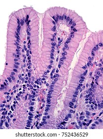
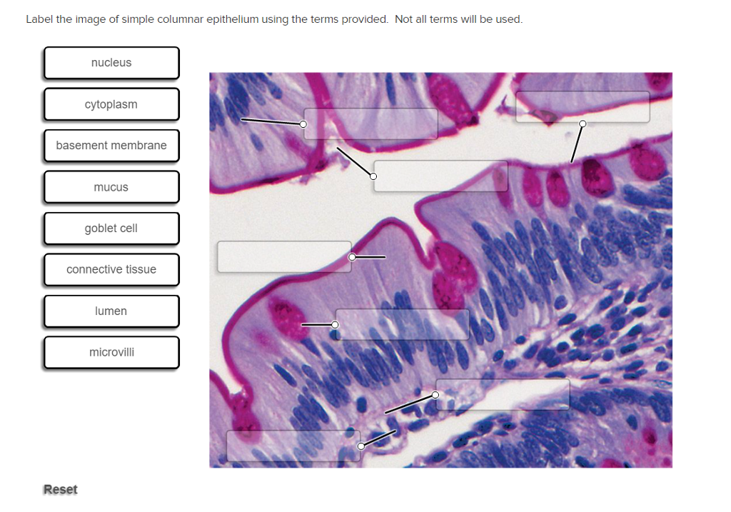
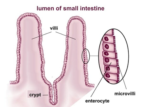
The mucosa is lined by simple columnar epithelium (lamina epithelialis) comprising enterocytes and goblet cells. Underneath lies a connective tissue layer (lamina propria) and a muscle layer (lamina muscularis mucosae). Compared to the rest of the small intestine the circular folds are rather flat and the villi relatively short.
Simple Columnar Epithelium: A Labeled Diagram and Functions Epithelium is a tissue that lines the internal surface of the body, as well as the internal organs. Simple epithelium is one of the types of epithelium that is divided into simple columnar epithelium, simple squamous epithelium, and simple cuboidal epithelium. Ciliated epithelium is a ...
Simple columnar cells cut tangentially to show how they form a very regular "pavement" when viewed from the surface. The cells are like tall blocks arranged very closely to each other with a small amount of tissue fluid in between. Slide 10 Detail of simple columnar epithelium with striated border (microvilli).
4 2 Epithelial Tissue Anatomy And Physiology. Simple Cuboidal Epithelium Tissue Body Tissues Basement. Simple Columnar Epithelium Basement Membrane. Stratified Cuboidal Epithelium Definition And Function Biology. Examining Epithelial Tissue Under The Microscope Human Anatomy. Lab Exercise 4 Epithelial Tissues Connective Tissue Proper What.
A. Simple columnar epithelium. Slide 29 (small intestine) View Virtual Slide Slide 176 40x (colon, H&E) View Virtual Slide Remember that epithelia line or cover surfaces. In slide 29 and slide 176, this type of epithelium lines the luminal (mucosal) surface of the small and large intestines, respectively. Refer to the diagram at the end of this chapter for the tissue orientation and consult ...

FIGURE 1-1 (a) Simple squamous epithelium lines the lumina of vessels, where it permits diffusion.(b) A photomicrograph of this tissue and (c) a labeled diagram.Simple squamous epithelia that line the lumina of vessels are referred to as endothelia, and that which cover visceral organs are referred to as mesothelia.

Epithelium is a tissue that lines the internal surface of the body, as well as the internal organs. Simple epithelium is one of the types of epithelium that is divided into simple columnar epithelium, simple squamous epithelium, and simple cuboidal epithelium. Bodytomy provides a labeled diagram to help you understand the structure and function of simple columnar epithelium.
The simple columnar epithelium is lining the villi and the mucosal surface of a duodenum. You will also find some goblet cells that intersperse among the absorptive columnar cells. The columnar absorptive cells have flattened nuclei located near the base and possess prominent microvilli that form a straight border.
Blood Vessels Labeled Simple - DIAGRAM OF HEART - Unmasa Dalha - Bodytomy provides a labeled diagram to help you understand the structure and function of simple columnar epithelium.. Hma practical 3 for monday july 23 and wednesday july 25. Capillaries are blood vessels that are one cell thick (endothelium) where the main diffusion and exchange ...

Simple columnar epithelium (100X) Primate small intestine At 100X you can begin to see the layer of simple columnar epithelium. the best place to look for it is on the surface of the villi. You can see what look like round white bubbles in the epithelium. These are goblet cells and they secrete mucus.

Start studying Simple Columnar Epithelium. Learn vocabulary, terms, and more with flashcards, games, and other study tools.
Simple Epithelium- it is composed of one layer of a cell and mostly has a secretory or an absorptive function. Compound (Stratified) Epithelium- it is made up of two or more than two layers of cells and mostly has a protective function. The glandular epithelium is made up of cuboidal or columnar cells. They are specialised for secretion.
The simple columnar epithelium is either ciliated or non-ciliated, and the non-ciliated columnar epithelium has microvilli on the apical area. Simple Columnar Epithelium Structure. The simple columnar epithelium is composed of a single layer of elongated cells that are always taller than they are wide and are linked directly to the basement ...


0 Response to "38 simple columnar epithelium labeled diagram"
Post a Comment