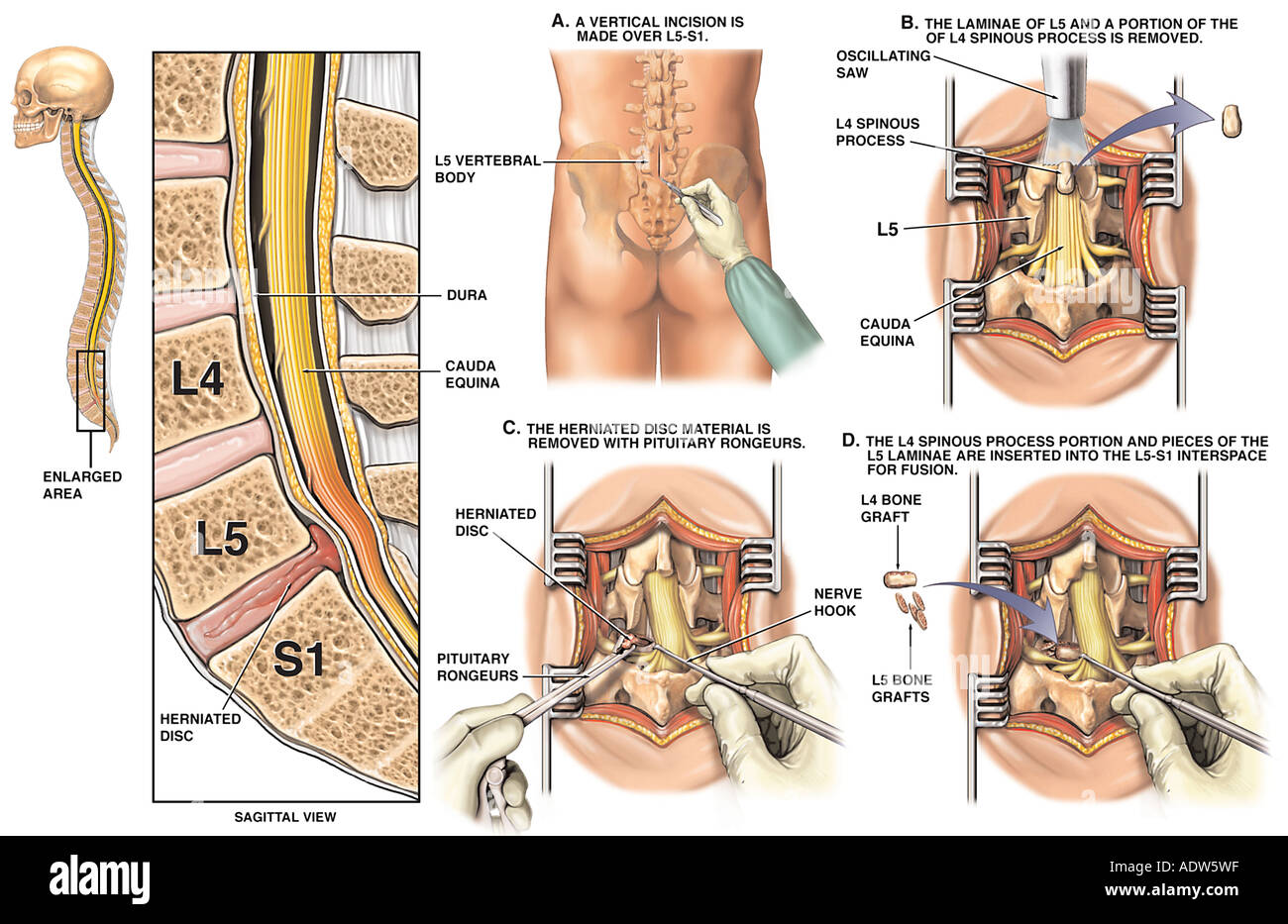
40 spine l5 s1 diagram
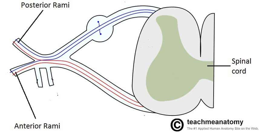
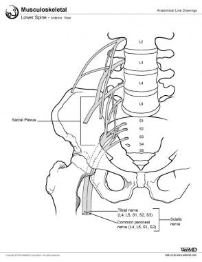
The plexus is formed by the anterior rami (divisions) of the sacral spinal nerves S1, S2, S3 and S4. It also receives contributions from the lumbar spinal nerves L4 and L5. In this article, we shall look at the anatomy of the sacral plexus - its formation and major branches. The fifth lumbar spine vertebrae (L5) is part of the greater lumbar region. To the human eye, this is the curve just above the buttocks, which is also commonly referred to as the small of the back ...
Spondylolisthesis: Diagram of L5 vertebra sitting correctly on the sacrum. Written by Mary Rodts, DNP. fig. 1. Diagram of an L5 vertebra "sitting" corrtectly on the sacrum. fig. 1a. Diagram of an L5 vertebra slipping forward on the sacrum (i.e., spondylolisthesis) Updated on: 02/01/10.

Spine l5 s1 diagram
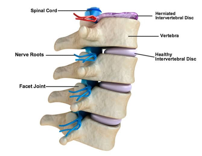
The five vertebrae of the lumbar spine are connected in the back by facet joints, which allow for forward and backward extension, as well as twisting movements. The two lowest segments in the lumbar spine, L5-S1 and L4-L5, carry the most weight and have the most movement, making the area prone to injury. L5 S1 Fusion refers to the level of the surgery. There are 5 spinal bones in the low back which are numbered from top to bottom L1, L2, L3, L4, and L5. Sandwiched between each of the spinal bones is a disc. The disc is named for the two spinal bones it is sandwiched between. For example, the lowest disc in the low back is the L5/S1 disc. (a) Diagram shows spinal fusion with a typical rod and screw device spanning the L4 through S1 vertebrae. (b) Photograph shows a metallic rod and screw device (Isola). (c, d) Anteroposterior (c) and lateral (d) radiographs show the same device as in b after positioning at the L4 through S1 vertebral levels. Radiopaque markers that delineate the ...
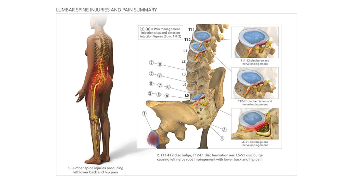
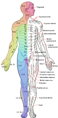
Spine l5 s1 diagram. Inferior Gemelli L5,S1 Quadratus Femoris L5,S1. NERVE TO OBTURATOR INTERNUS Superior Gemelli L5,S1 Obturator Internus L5,S1. NERVE TO PIRIFORMIS Piriformis L(5),S1,2. Muscle Innervation Chart: Head, Neck and Trunk. GREATER OCCIPITAL N. (POST. PRIMARY RAMI) Lower Back Pain. Back pain is a common symptom of an L5-S1 degenerative disc. The pain is usually located in the midline of the lower back. It is generally a chronic, mild to moderate aching sensation, with intermittent flare-ups of severe pain lasting for a few days or weeks.. Back pain from a degenerative disc is typically worse with sitting, bending, twisting, sneezing or coughing. changes at L5 / S1 showing signs of fusion spondylolisthesis at L4 / 5 lumbar vertebra slipped forward degenerative changes in facet joint nerve root being compressed Diagram 1 showing spondylolisthesis at L4 / L5 X-ray showing spondylolisthesis at L4 / L5 L4 L5 L3 S1 L3 L4 L5 S1 When nerves are compressed they can produce symptoms of pain, 740 lumbar spine anatomy diagram stock photos, vectors, and illustrations are available royalty-free. See lumbar spine anatomy diagram stock video clips. of 8. spinal vertebrae bone spine vertebra toracica spinal cord spine structure back diagram spine sections spinal cord vertebrae spinal structure health diagram. Try these curated collections.
File under medical illustrations showing L5-S1 Disc Space, with emphasis on the terms related to medical disc space spine l5 s1 intervertebral nerve root spinal nerves dura epidural ligamentum flavum. This medical image is intended for use in medical malpractice and personal injury litigation concerning L5-S1 Disc Space. Examination. She presents with isolated back pain over the L4-L5 and L5-S1 facet joints. Flexion is 60% of normal. Extension is limited to 20% of normal because of severe back pain. Straight leg raising tests are bilaterally negative. Strength is normal, but slightly diminished right to L5 light-touch sensation. Lumbar interbody fusion: A degenerated disc is removed and L5-S1 vertebrae are fused together with implants or bone grafts. While performing a fusion surgery, the spinal fixation of the S1 segment usually presents a greater risk of failure (pseudarthrosis) compared to L5. To avoid this complication, the addition of an interbody support (device ... Back pain or leg pain can typically arise due to injury where the lumbar spine and sacral region connect (at L5 - S1) because this section of the spine is subjected to a large amount of stress and twisting. People with rheumatoid arthritis or osteoporosis are inclined to develop stress fractures and fatigue fractures in the sacrum.
OSMOSIS.ORG 709. This Osmosis High-Yield Note provides an overview of Spinal disorders essentials. All Osmosis Notes are clearly laid-out and contain striking images, tables, and diagrams to help visual learners understand complex topics quickly and efficiently. Find more information about Spinal disorders by visiting the associated Learn Page. The L5 S1 disc in particular is the most fragile and susceptible to protrusion since it often carries more weight than the other lumbar discs. ((L5 is medical shorthand for the fifth vertebrae in the lumbar, or the lower part of the spine, and S1 denotes the first vertebrae in the sacrum. The L5 S1 disc is sandwiched between these two vertebrae). The lumbar spine receives a high degree of stress. Watch: Lumbar Spine Anatomy Video The L5-S1 motion segment has distinctive anatomy and receives a higher degree of mechanical stress and loads compared to the segments above. 1 These characteristics may make L5-S1 susceptible to traumatic injuries, degeneration, disc herniation, and/or nerve pain. L5-S1 is the exact spot where the lumbar spine ends and the sacral spine begins. The lumbosacral joint is the joint that connects these bones. L5-S1 is composed of the last bone in the low back, called L5, and the triangle-shaped bone beneath, known as the sacrum. The sacrum is made of five fused bones, of which the S1 is the topmost.
Listed are some commonly occurring changes, which may be described in a lumbar MRI report. The most common levels affected at L4-5 and L5-S1 as these are the most mobile levels in the lumbar spine. About 60% of a person's range of motion is at L4-5 and L5-S1. Lumbar Spondylosis
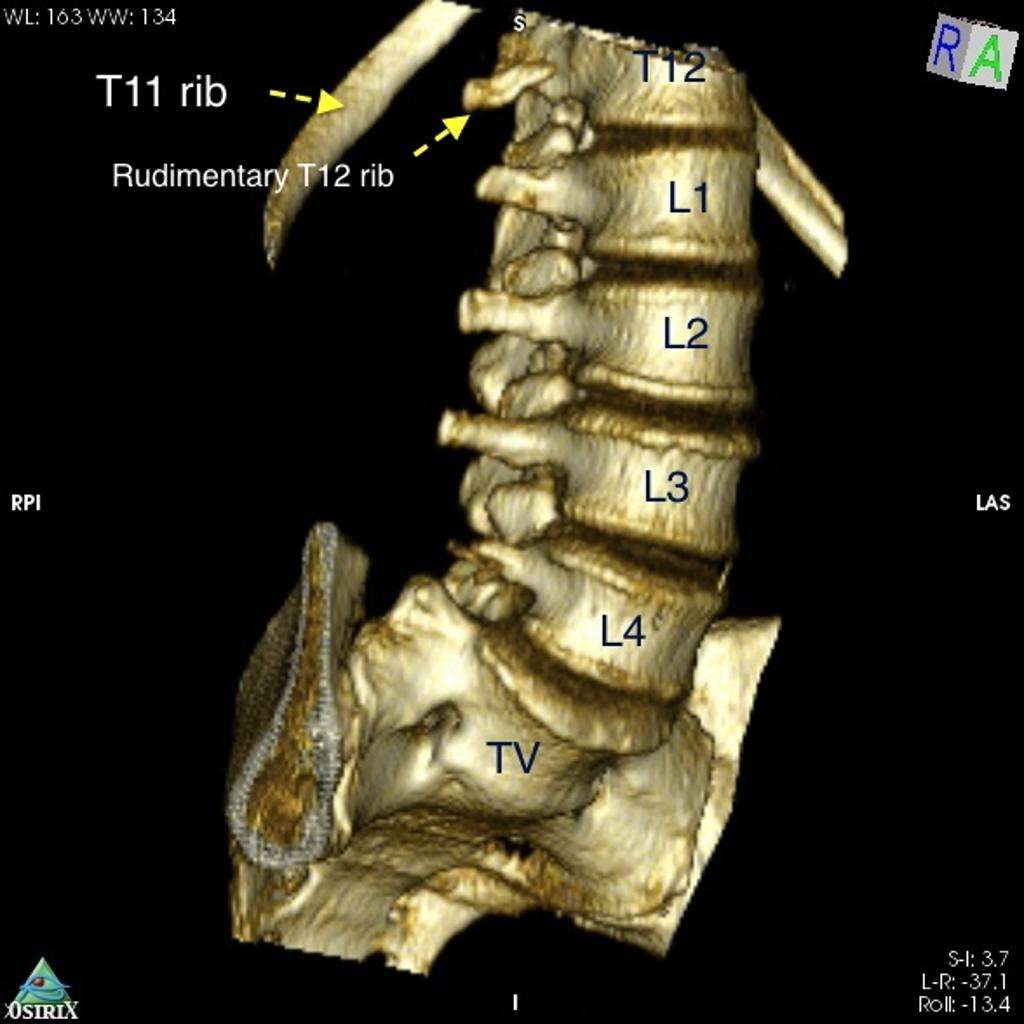
The L4 and L5 are the two lowest vertebrae of the lumbar spine. Together with the intervertebral disc, joints, nerves, and soft tissues, the L4-L5 spinal motion segment provides a variety of functions, including supporting the upper body and allowing trunk motion in multiple directions. 1. Lumbar Spine Anatomy Video Save.
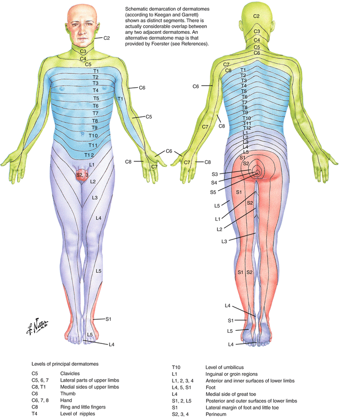
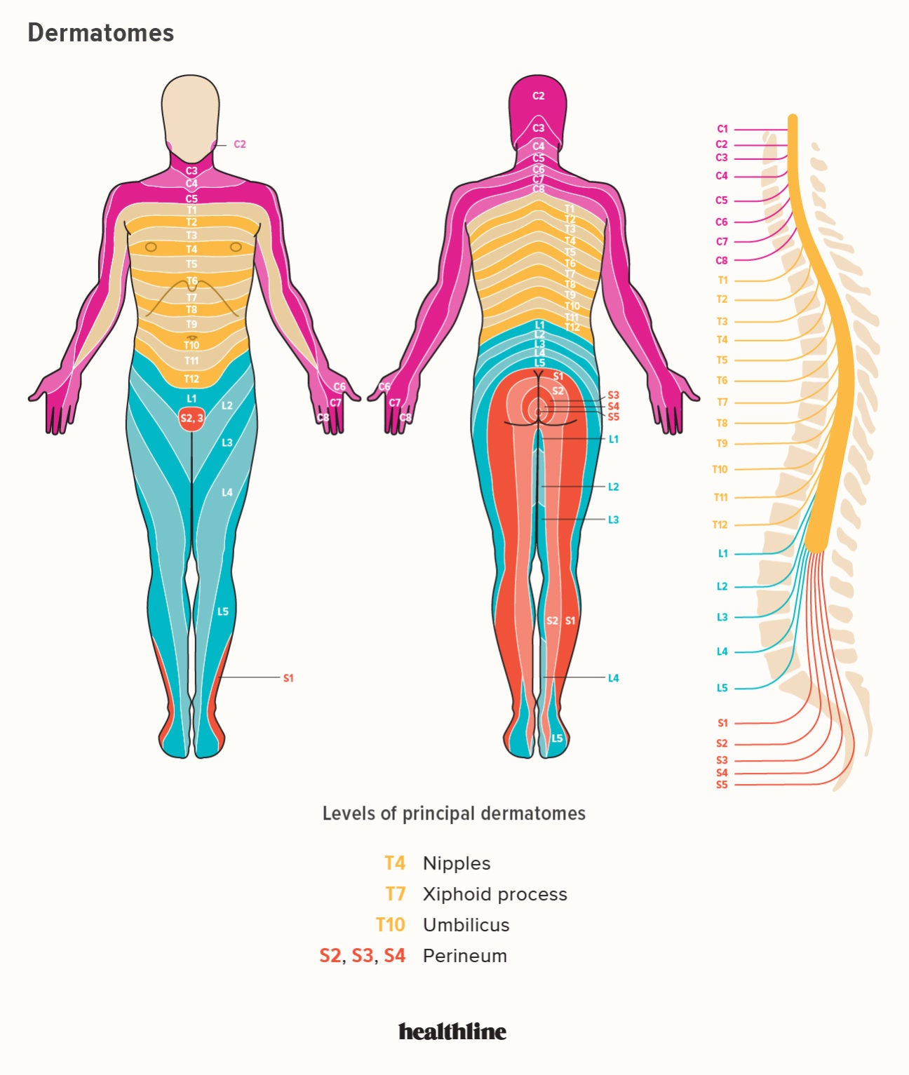
Pain. Pain is a common symptom associated with L5-S1 pinched nerves 3.This may feel like a dull ache or a sharp pain. L5 nerve compression causes pain along the outer border of the back of your thigh, while S1 nerve compression causes pain in your calf and the bottom of your foot 3. This pain can range from mild to severe and may be constant or intermittent.
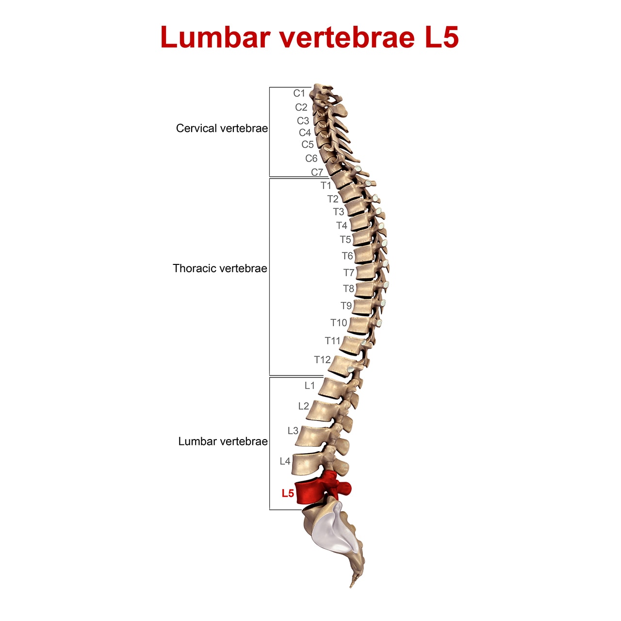
The lumbar spine is located in the lower back below the cervical and thoracic sections of the spine. It consists of five vertebrae known as L1 - L5. These lumbar vertebrae (or lumbar bones) contain spinal cord tissue and nerves which control communication between the brain and the legs. Damage to the lumbar spinal cord subsequently affects the ...
The sacral spine consists of five segments, S1 - S5, that together affect nerve communication to the lower portion of the body. It is important to understand that the spinal cord does not extend beyond the lumbar spine. L2 is the lowest vertebral segment that contains spinal cord. After that point, nerve roots exit each of the remaining ...
Spinous processes of L5,L4,L3 and the caudal one-third of L2 removed with Leksell rongeur. The laminae of L5,L4, and L3 thinned down with Leksell ronguer. Utilizing a curette, caudal one-third of L5 lamina undercut with curette, and bilateral L5 laminectomy performed with Leksell and Kerrison ronguers. Ligmentum flavum between L5 - S1, L4
Spinal Cord Diagram at the Canadian Paraplegic Association (NS) web site. Spinal Cord Anatomy Apparelyzed.com. This series of eight guides describes outcomes according to level of spinal cord injury (C1-3, C4, C5, C6, C7-8, T1-9, T10-L1 and L2-S5). Each guide provides individual guidance on what people with different levels of SCI can ...
The most common levels in the spine requiring treatment are L4-5 and L5-S1 fusion. Many patients requiring lumbar fusion surgery also have pinched nerves from herniated discs or spinal stenosis. As a result, the surgery is often performed in conjunction with microlumbar discectomy or lumbar laminectomy .
Spine Diagrams. The human spine consists of 33 vertebrae: 7 Cervical vertebra (C1-C7) 12 Thoracic vertebra (T1-T12) 5 Lumbar vertebra (L1-L5) The sacrum and coccyx are made up of 9 fused vertebrae. Each vertebra is attached to the one above and below it by ligaments and muscles.
Impingement of a nerve between the L5 and S1 vertebrae indicates the structure is placing pressure on the nerve root. According to the Laser Spine Institute, this is one of the most common of all pinched nerves. This nerve root feeds the sciatic nerve, and impingement has the potential to affect the lower buttocks, legs and feet.
Diagram of muscle attachments at the L4/5 and L5/S1 levels. The separate muscle bundles at L4/5 are replaced at the L5/S1 level by a fascial sheet or aponeurosis that extends laterally over the ...
(a) Diagram shows spinal fusion with a typical rod and screw device spanning the L4 through S1 vertebrae. (b) Photograph shows a metallic rod and screw device (Isola). (c, d) Anteroposterior (c) and lateral (d) radiographs show the same device as in b after positioning at the L4 through S1 vertebral levels. Radiopaque markers that delineate the ...
L5 S1 Fusion refers to the level of the surgery. There are 5 spinal bones in the low back which are numbered from top to bottom L1, L2, L3, L4, and L5. Sandwiched between each of the spinal bones is a disc. The disc is named for the two spinal bones it is sandwiched between. For example, the lowest disc in the low back is the L5/S1 disc.
The five vertebrae of the lumbar spine are connected in the back by facet joints, which allow for forward and backward extension, as well as twisting movements. The two lowest segments in the lumbar spine, L5-S1 and L4-L5, carry the most weight and have the most movement, making the area prone to injury.



:max_bytes(150000):strip_icc()/human-spine--pelvis--chiropractic--orthopedic--medical-model--heathcare--isolated-157403981-5bfd7bd9c9e77c0051d77683.jpg)





:background_color(FFFFFF):format(jpeg)/images/library/12522/spine-bones-and-ligaments-Recovered_english.jpg)






0 Response to "40 spine l5 s1 diagram"
Post a Comment