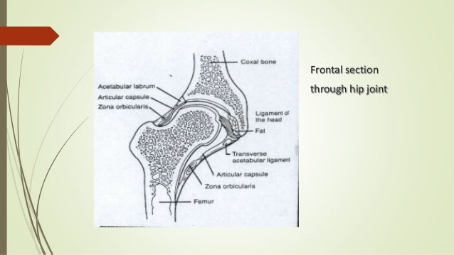
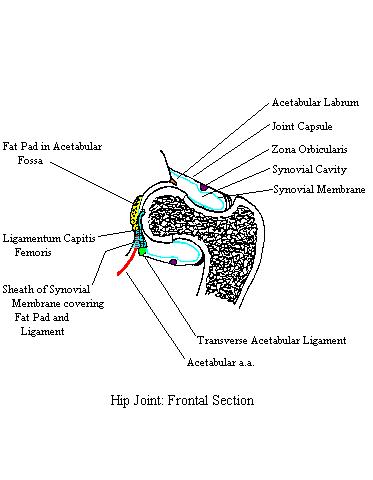
35 the diagram shows a frontal section of the hip joint
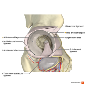
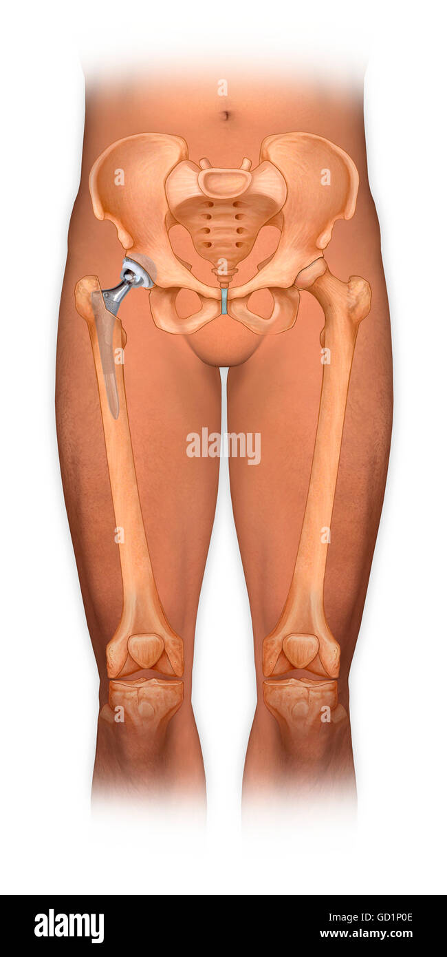
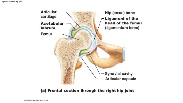
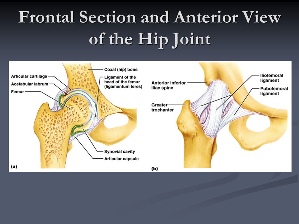
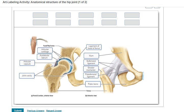
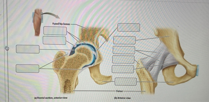
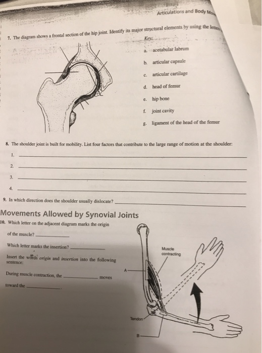
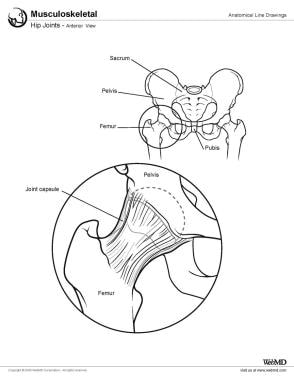
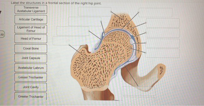
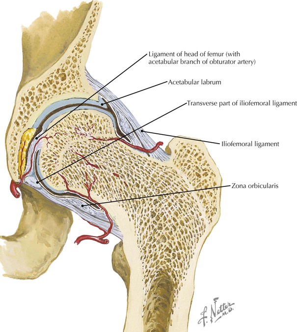
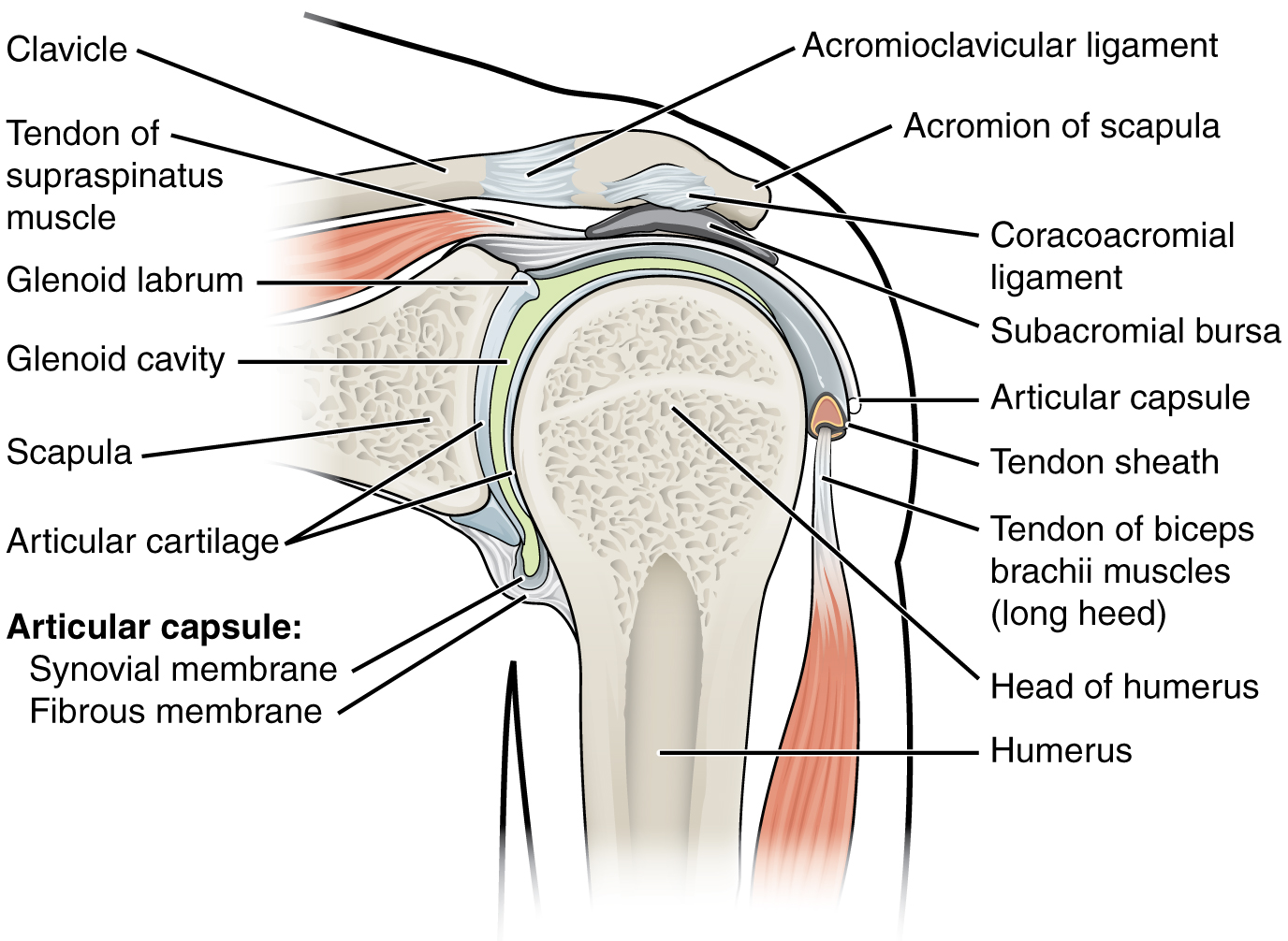
Jun 07, 2017 · The diagram on the right shows a cross section of the hip. As you can see, the top of the femur is shaped like a ball and the concave cavity of the pelvis is shaped like a socket. Appropriately named, the hip is a ball and socket joint. The round head of the femur spins and moves inside the deep socket of the pelvis. The diagram shows a frontal section of the hip joint. Identify its major structural elements by using the key letters. Key: a. acetabular labrum b. articular capsule c. articular cartilage d. coxal bone e. head of femur f. ligament of the head of the femur g. synovial cavity 8. The shoulder joint is built for mobility. List four
View Pearson eText15 from BIO 201 at Pima County Community College. Review Sheet 11 183 7. The diagram shows a frontal section of the hip joint.
The diagram shows a frontal section of the hip joint
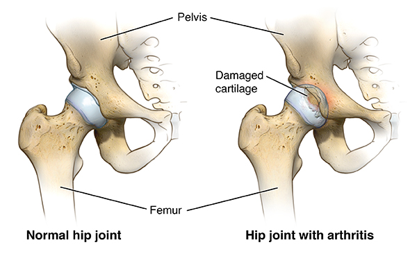
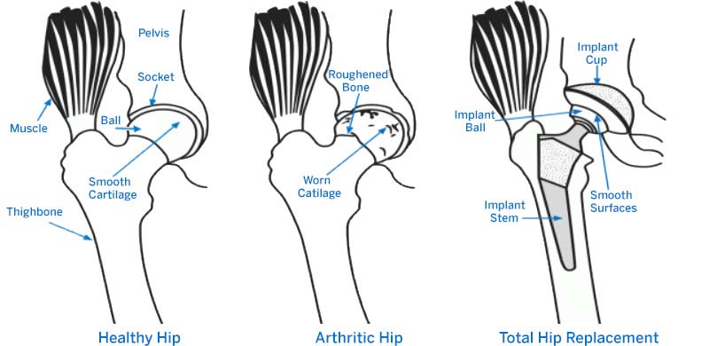
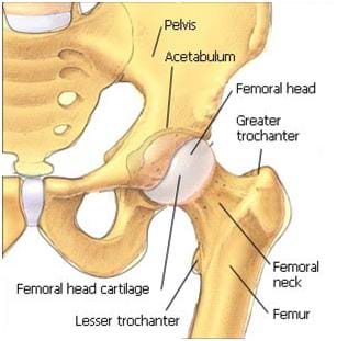
7. The diagram shows a frontal section of the hip joint. Identify its major structural elements by using the key letters. Normally, a smooth cushion of shiny white hyaline (or articular) cartilage about 1/4 inch thick covers the femoral head and the acetabulum. The articular cartilage is kept slick by fluid made in the synovial membrane (joint lining). Synovial fluid and articular cartilage are a very slippery combination—3 times more slippery than skating on ice and 4 to 10 times more slippery than a metal on plastic hip replacement. Synovial fluid is what allows us to flex our joints under great pressure without wear. Since the cartilage is smooth and slippery, the bones move against each other easily and without pain. When the cartilage is damaged, whether secondary to osteoarthritis (wear-and-tear type arthritis) or trauma, joint motion can become painful and limited. The hip joint is one of the largest joints in the body and is a major weight-bearing joint. Weight bearing stresses on the hip during walking can be 5 times a person’s body weight. A healthy hip can support your weight and allow you t... Author's Note: I made this as a kinda Wikipedia style biological summary of Valfalk/Valphalk/Valstrax with speculative features and explanations for its abilities. Do note that this is heavily speculative and mostly based on my very basic understanding of biology and general observation with these monsters. It is all labelled so you can skip to specific parts if you want. However, I put a lot of effort into this one in particular (Almost double the word count on the Zinogre post) and I’d recomme...
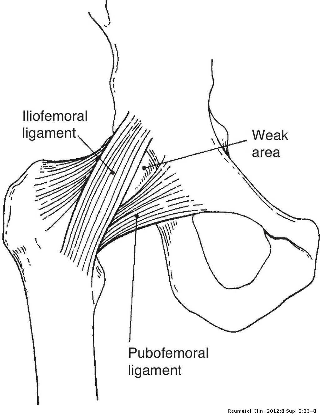
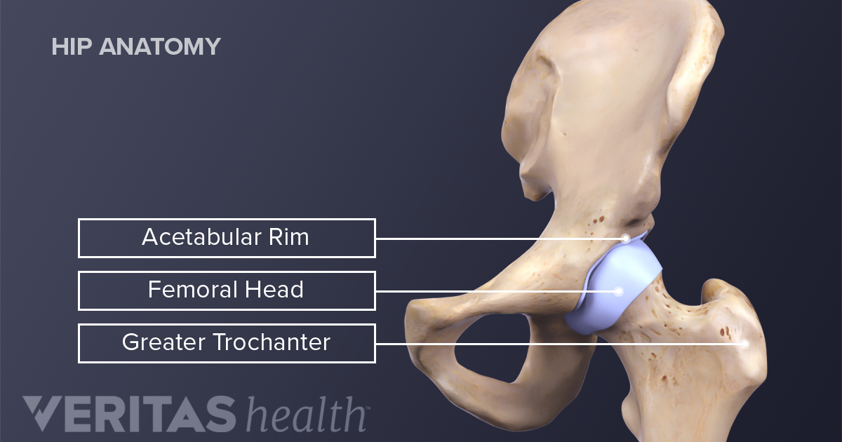
The diagram shows a frontal section of the hip joint. Start studying hip joint frontal section. Learn vocabulary, terms, and more with flashcards, games, and other study tools. Cross post from r/gratefuldoe OK, so truly a collision of worlds, with deadheads reaching out to the Zombies for help, but here we go! A young man was found after an apparent hit and run July 20th 1995, near Atlanta GA. The location where he was found was with a few miles and on the same road as a White Zombie, Kyuss, Babes in Toy Land, Reverend Horton Heat show at the Lakewood Amphitheater. He had a few tattoos including one on his hand (rarer in the 90's). It is not known if he attended ... Jan 01, 2019 · The hip is a ball-and-socket joint, similar to the joint in the shoulder. Part of the reason for the hip’s stability is that there is a very deep socket, called the acetabulum, in the hip joint. A strong capsule joint supported by ligaments and muscles also provides extra stability to the hip. The diagram shows a frontal section of the hip joint. Identity its major structural elements by using the letters Key: a. acetabular labrum b. articular ...
The diagram shows a frontal section of the hip joint. Identity its major structural elements by using the letters Key: a. acetabular labrum b. articular capsule c. articular cartilage d. head of femur e. hip bone f. joint cavity g. ligament of the bead of the femur The shoulder joint is built for mobility. List four factors that contribute to the large range of motion at the shoulder: 1. Right hip joint. Posterior half, viewed from in front. The joint surfaces have been somewhat pulled apart. Author's Note: I made this as a kinda Wikipedia style biological summary of Valfalk/Valphalk/Valstrax with speculative features and explanations for its abilities. Do note that this is heavily speculative and mostly based on my very basic understanding of biology and general observation with these monsters. It is all labelled so you can skip to specific parts if you want. However, I put a lot of effort into this one in particular (Almost double the word count on the Zinogre post) and I’d recomme... Normally, a smooth cushion of shiny white hyaline (or articular) cartilage about 1/4 inch thick covers the femoral head and the acetabulum. The articular cartilage is kept slick by fluid made in the synovial membrane (joint lining). Synovial fluid and articular cartilage are a very slippery combination—3 times more slippery than skating on ice and 4 to 10 times more slippery than a metal on plastic hip replacement. Synovial fluid is what allows us to flex our joints under great pressure without wear. Since the cartilage is smooth and slippery, the bones move against each other easily and without pain. When the cartilage is damaged, whether secondary to osteoarthritis (wear-and-tear type arthritis) or trauma, joint motion can become painful and limited. The hip joint is one of the largest joints in the body and is a major weight-bearing joint. Weight bearing stresses on the hip during walking can be 5 times a person’s body weight. A healthy hip can support your weight and allow you t...
7. The diagram shows a frontal section of the hip joint. Identify its major structural elements by using the key letters.
Hip Anatomy Physiopedia

Image Jpg Articulations And Body Movements 7 The Diagram Shows A Frontal Section Of The Hip Joint Identify Its Major Structural Elements By Using The Course Hero

Elbow Joint Open Educational Resource Oer Unsyiah Library
Anatomy Hip Joint Diagram High Resolution Stock Photography And Images Alamy
Biomechanics Of Hip
8 5 No Vids

11187 Jpeg Review Sheet 11 187 7 The Diagram Shows A Frontal Section Of The Hip Joint Identify Its Major Structural Elements By Using The Key Letters Course Hero
Articulations A Look At The Structural And Functional Classification Of Joints And The Movements They Provide Ppt Video Online Download
Hip Joint Seen From Before Clipart Etc

Hip Pain Explained Including Structures Anatomy Of The Hip And Pelvis
Art Labeling Activity Anatomical Structure Of The Chegg Com

Stick Diagram Showing The Analysed Hip And Knee Angles In The Download Scientific Diagram
Solved Rused Hip Bones 10 Femur A Frontal Section Anterior Chegg Com
1

Regional Biomechanics Hip Joint Ppt Video Online Download

Hip Thigh Atlas Of Anatomy
Solved The Diagram Shows A Frontal Section Of The Hip Joint Chegg Com
Clinical Anatomy Of The Pelvis And Hip Reumatologia Clinica
Hip Replacement Surgery Johns Hopkins Medicine
Hip Joint Diagram High Resolution Stock Photography And Images Alamy
Reduction Of Posterior Hip Dislocation Background Indications Contraindications

Uk Advice For Gp S Hip Joint Replacements Earl S View
Hip Joint Concise Medical Knowledge

Coronal Plane Wikipedia
Hip Replacement Surgery Procedure Types And Risks Hss

Joints Joint Movements Ppt Video Online Download
Hipjointfrontalcomplete
Solved Label The Structures In A Frontal Section Of The Chegg Com

Anterior Hip Pain Pain At The Front Of The Hip
Acetabulum
Hip Anatomy
Hip Joint Diagram High Resolution Stock Photography And Images Alamy
Why Does The Front Of My Hip Pinch
Lower Limb Radiology Key
2 2 4 Anatomy Of Selected Synovial Joints Biomechanics Of Human Movement
0 Response to "35 the diagram shows a frontal section of the hip joint"
Post a Comment