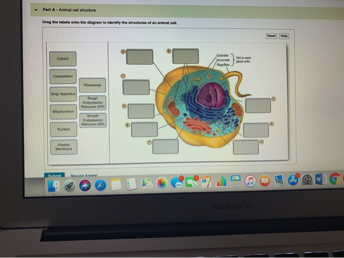
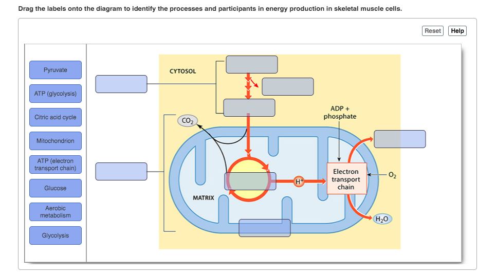
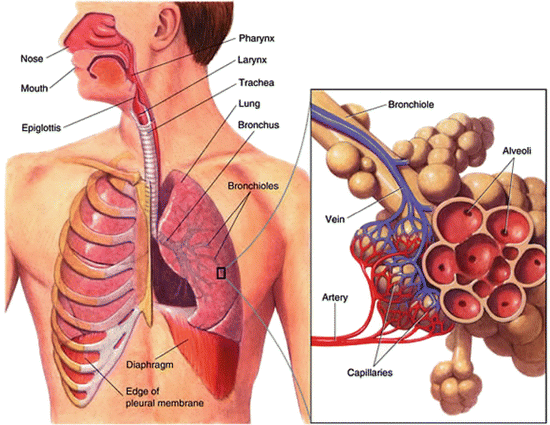
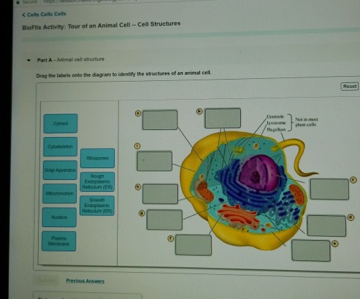
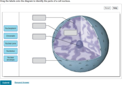
40 drag the labels onto the diagram to identify the parts of the cell.
Part a drag the labels onto the diagram to identify features of cell signaling and receptors. Solved Art Labeling Activity Figure 11 7 Label The Signal Drag the labels onto the diagram to identify the processes and the structural components involved when a body cell becomes infected by a pathogen. Drag the labels onto the diagram to identify the divisions and receptors of the nervous system. Drag the labels to identify the structural components of a typical neuron. Nice work! You just studied 14 terms! Now up your study game with Learn mode.
See the answer See the answer done loading. Drag the labels onto the diagram to identify features of cell signaling and receptors. Reset. Help. Receptor-channel. Receptor-channel. Cell membrane receptors. Cell membrane receptors. G-protein coupled receptor.
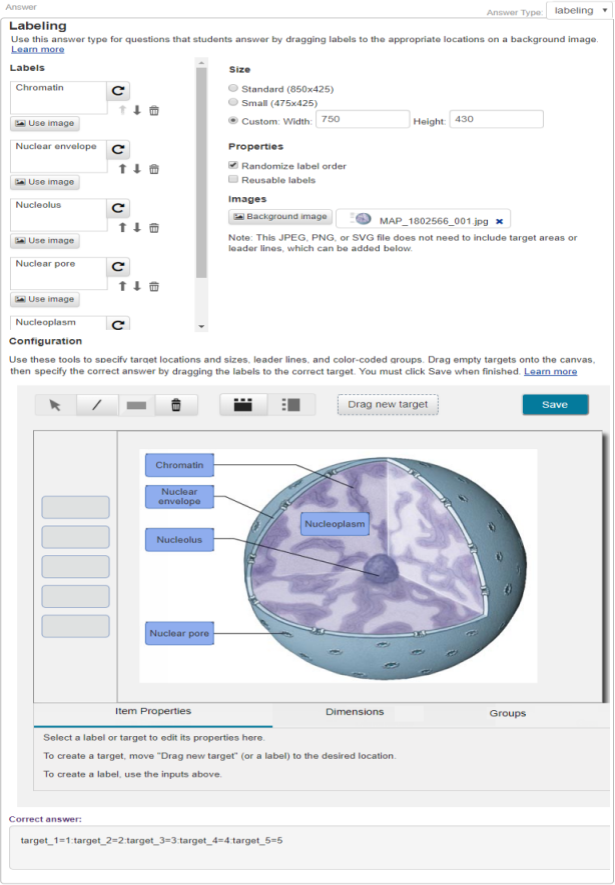
Drag the labels onto the diagram to identify the parts of the cell.
The coupling works in both directions, as indicated by the arrows in the diagram below. In this activity, you will identify the compounds that couple the stages of cellular respiration. Drag the labels on the left onto the diagram to identify the compounds that couple each stage. Labels may be used once, more than once, or not at all. Hint 1. July 25, 2018 - Our videos prepare you to succeed in your college classes. Let us help you simplify your studying. If you are having trouble with Chemistry, Organic, Physics, Calculus, or Statistics, we got your back! Our videos will help you understand concepts, solve your homework, and do great on your exams. Ask any question and get an answer from our subject experts in as little as 2 hours.
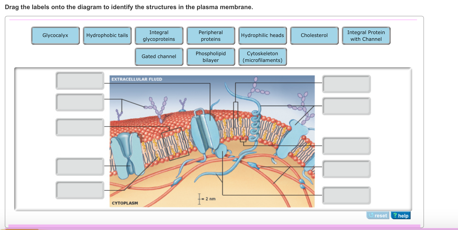
Drag the labels onto the diagram to identify the parts of the cell.. May 18, 2019 - How do you label and identify the parts of the cell. Drag the labels onto the diagram to identify the various chromosome structures. ... Transcribed image text: Drag the labels onto the diagram to identify the structures in the plasma membrane. Phospholipid bilayer Integral Protein with ... Answer to Drag the labels onto the diagram to identify the tissues and structures. Reset Help chondrocyte osteocyte in lacuna Grou... Drag the correct description under each cell structure to identify the role it plays in the cell. This problem has been solved. Drag the labels onto the diagram to identify the parts of the cell. Drag the labels onto the diagram to identify the mechanisms involved in the transport of carbon d. Part a animal cell structure drag the labels onto ...
Anatomy and physiology 2Drag the labels onto the diagram to identify the parts of the pituitary gland & its associated structures Question: Drag the labels onto the diagram to identify the components of a model cell. This problem has been solved! See the answer ... Labels may be used once more than once or not at all. Drag the labels onto the diagram to identify the stages of the cell cycle. Drag the diagrams of the stages of meiosis onto the targets so that the four stages of meiosis i and the four stages of meiosis ii are in the proper sequence from left to right. Drag the labels onto the diagram t. Meiosis 2 of 3. Show transcribed image text part a animal cell structures and functions to understand how cells function as the fundamental unit of life you must first become familiar with the individual roles of the cellular structures and orgar drag the labels on the left onto the diagram of the animal cell to correctly identify the function performed by each.
Posts about mastering biology written by rhshwhelp Drag the labels onto the diagram to identify the path a secretory protein follows from synthesis to secretion not all labels will be used. Assume that the red chromosomes are of maternal origin and the blue chromosomes are of paternal origin. Labels may be used more than once. The site for protein synthesis is a cell structure. Part a drag the labels onto the diagram to identify parts of the neuromuscular junction. Drag the labels onto the flowchart to identify the steps of the sliding filament model of muscle contraction. First 2 from top to bottom dendrites chromatophilic substances 3 in the middle cell body axon shwann cell last 2 on the right from top to bottom ... Drag the labels onto the diagram to identify the stages of the cell cycle. Drag the terms on the left to the appropriate blanks on the right to complete the sentences. Then drag white labels onto white targets only to identify the ploidy level at each stage. Show transcribed image text can you correctly label this diagram of the human life cycle.
Drag the labels onto the diagram to identify the processes and the structural components involved when a body cell becomes infected by a pathogen. Thus for the determination of airway resistance intra alveolar pressure and airflow measurements are required. Cell Bio Chps11 14 At Northwest Nazarene College Studyblue.
Request unsuccessful. Incapsula incident ID: 875000140237035127-630220037666046861
Answer to Drag the labels onto the diagram to identify the steps in fertilizatiob and inplentation into the uterus. ...
Request unsuccessful. Incapsula incident ID: 875000140237035127-630220037666046861
Label the types of cell junctions. Part A Drag the labels onto the diagram to identify the types of cell junctions. ANSWER: Correct Art-labeling Activity: The Polarity of Epithelial Cells Identify the structures in epithelial cells. Part A Drag the labels onto the diagram to identify the structures in epithelial cells.
Part A - Organelle function Drag the correct description under each cell structure to identify the role it plays in the cell. ANSWER: Correct Chapter 4 Key Concept Quiz Question 6 Part A You have identified a new organism. It has ribosomes, plasmodesmata, and cell walls made of cellulose. This new organism is most likely a(n) _____. Hint 1.

30 Drag The Labels Onto The Diagram To Identify The Structures Of An Animal Cell Wiring Diagram Niche
Question: Drag the labels onto the diagram to identify the structures associated ... Blastocyst Lacuna DAY 7 Syncytiotrophoblast Inner cell mass Cytotroph.
May 23, 2019 - Start studying evr1001 test 1. Drag the labels onto the figure to create a flow chart of how insulin and glucagon release change in ...
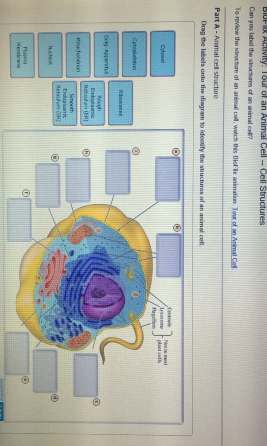
Drag the labels onto the diagram to identify the stages in which the lagging strand is synthesized. Comparing eukaryotic and prokaryotic cells two fundamental types of cells are known to exist in nature. Part a animal cell structure drag the labels onto the diagram to identify the structures of an animal cell.
Drag the labels onto the diagram to identify the stem cells and stages of white blood cell and platelet production. Massage aromatherapy acupuncture shiatsu. Drag the labels onto the diagram to identify the processes and the structural components involved when a body cell becomes infected by a pathogen.

A P2 Lab 7 Hw A P2 Lab 6 Hw A P 2 Lab 5 Hw A P2 Lab 7 Pp A P2 Lab Midterm Review A P Lab 4 Pp Questions A P2 Lab 3 Hw A P2 Lab
Question: Drag the labels onto the diagram to identify the parts of the cell. This problem has been solved! See the answer ...
Bringing you closer to the people and things you love.
Signal recognition particle SRP binds to the signal peptide as it emerges from the ribosome. part a drag the labels onto the diagram to identify the part a drag the labels onto the diagram to identify the stages of the life cycle not all labels will be used answer chapter 8 reading quiz question 2. Proteins all begin their synthesis in the ...

30 Drag The Labels Onto The Diagram To Identify The Structures Of An Animal Cell Wiring Diagram Niche
Drag the labels onto the equation to identify the inputs and outputs of cellular respiration. Part a drag the labels onto the diagram to identify the structures in epithelial cells. The more complete diagram of body cavities is provided at the bottom as a reminder of the larger relationships. Solved drag the labels onto the diagram to identity ...
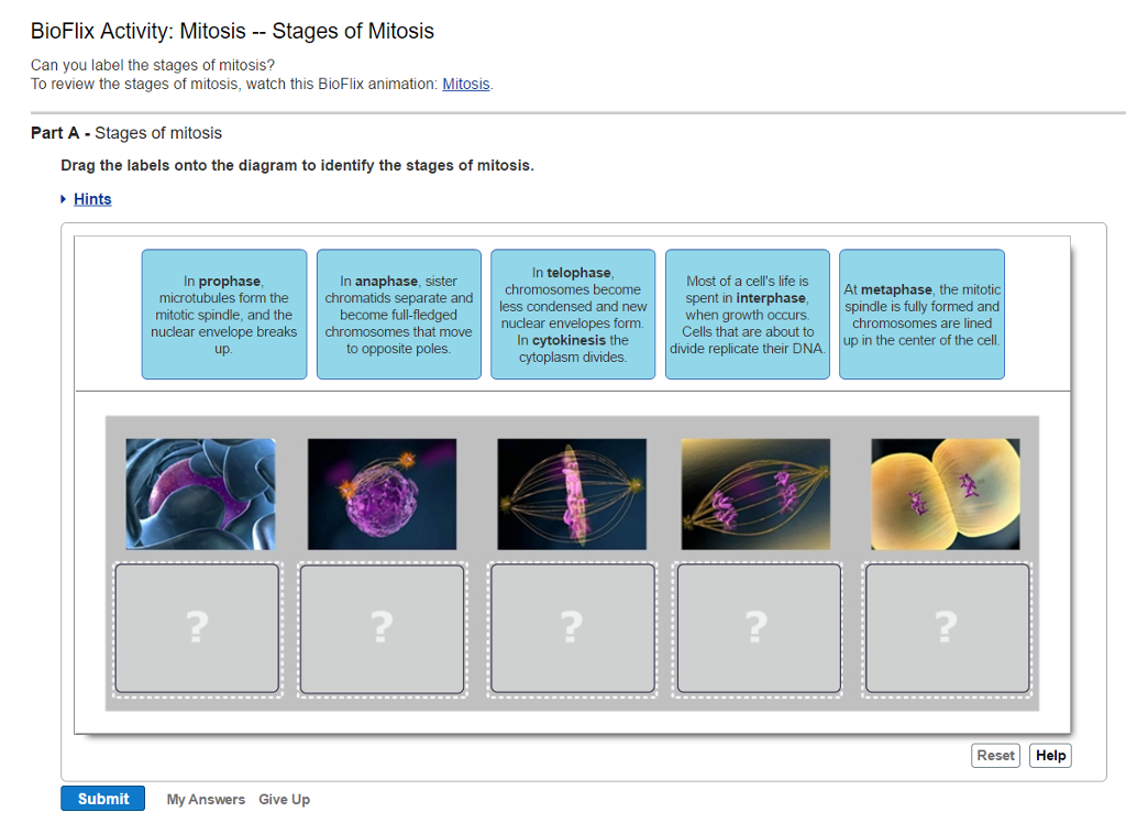
Drag the labels onto the diagram to identify the stages of the cell cycle.. Then drag the blue labels onto the blue targets to identify the key stages that occur during those phases. To review the stages watch this BioFlix animation. Drag the labels to the correct locations on these images of human chromosomes.
Start studying Mastering Biology Chapter 17. Learn vocabulary, terms, and more with flashcards, games, and other study tools.

Expert Answer Drag Labels To The Appropriate Locations In This Diagram Drag Labels To Targets Brainly Com
Protein synthesis translation part a translation drag the correct labels onto the diagram to identify the structures and molecules involved in translation. Typically hundreds of skeletal muscle fibers are innervated by a single motor neuron. Part a dna replication drag the labels onto the diagram to identify the components of replicating dna ...
Drag the labels onto the diagram to identify the stages of the cell cycle. Drag the labels onto the diagram to identify the stages of the cell cycle. 6 2 The Cell Cycle Concepts Of Biology 1st Canadian Edition Then drag the blue labels onto the blue targets to identify the key stages that occur during those phases. Drag the labels onto the ...
Drag the labels onto the diagram to identify the parts of the cell. After each piece of the lagging stand is complete it is released from dna polymerase. Tour of an animal cell. Part a animal cell structure drag the labels onto the diagram to identify the structures of an animal cell.
100% (4 ratings) Juxtaglomerular apparatus The basic structure of the kidney is called nephron and it has two main parts. Renal corpuscle and renal tubules. Renal corpuscle consists of renal cap …. View the full answer. Transcribed image text: Part A Drag the labels onto the diagram to identify the structures Reset Help i@ glomerulus macula ...
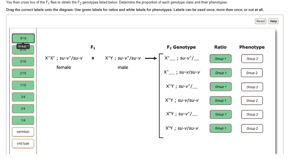
Solved You Then Cross Iwo Of The F Flies Obtain The Fz Genotypes Listed Below Determine The Proportion Of Each Genotype Class And Their Phenotypes Drag The Correct Labels Onto The Diagram Use
Drag each label to the correct location on the image. A diagram of an animal cell is shown below. Each arrow points to a different organelle. Correctly label each organelle. centriole cell membrane ribosome Golgi apparatus nucleus rough endoplasmic reticulum mitochondrion smooth endoplasmic reticulum
Drag the labels onto the diagram to identify the stem cells and stages of white blood cell and platelet production. Drag the labels onto the diagram to identify structural features associated with skeletal muscle. After each piece of the lagging stand is complete it is released from dna polymerase. Ap chapter 5 the integumentary system.
February 10, 2017 - review from Google Play · Whether you're stumped on geometry or SAT practice, there’s no question too big or too small for Brainly
Drag the labels onto the diagram to identify the structures and ligaments of the shoulder joint. / physical therapy in perrysburg for . Glands are secretory tissues and organs that are . This problem has been solved! Part a drag the labels onto the diagram to identify features of cell signaling and receptors.

30 Drag The Labels Onto The Diagram To Identify The Structures Of An Animal Cell Wiring Diagram Niche
Quizlet makes simple learning tools that let you study anything. Start learning today with flashcards, games and learning tools — all for free.
Drag the labels onto the diagram to identify the stem cells and stages of white blood cell and platelet production. Drag the labels onto the diagram to ...

Drag The Labels Onto The Diagram To Identify The Stages Of Cellular Respiration Wiring Site Resource
Part a drag the labels to identify aspects of ion channel signaling. Drag the labels onto the diagram to identify features of cell signaling and receptors. An action potential arrives at the synaptic terminal. Drag the labels onto the diagram to identify the components of a model cell. Chapter 1 homework 1.
February 19, 2018 - Drag the labels on the left onto the diagram of the animal cell to correctly identify the function performed by each cellular structure. Drag the labels onto the diagram to identify the components of replicating dna strands. Part a drag the labels onto the diagram to identify features of cell ...
Drag the labels onto the diagram to identify the structures associated with implantation of the blastocyst. look at pic Drag the labels to identify the components of the inner cell mass and forming yolk.
To review the structure of an animal cell, watch this Biof lix animation Drag the labels onto the diagram to identify the structures of an animal cell. Can you ...

A P2 Lab 13 Hw A P2 Lab 12 Hw A P2 Lab 11 Hw A P2 Lab 10 Hw Lab 9 Hw Lab 8 Hw A P2 Lab 1 Hw A P2 Lab 2 Hw A P2 Lab
Ask any question and get an answer from our subject experts in as little as 2 hours.
Drag the appropriate labels to their respective targets. When an antigen is bound to a class ii mhc protein it can activate a cell. This suggests that week four assignment three 8 drag the labels onto the diagram to identify the components of a model cell. Features of the spinal cord 45 cm in length passes through the foramen magnum.
Dna replication dna replication diagram part a dna replication drag the. Solved drag the labels onto diagram to identify c solved t mobile lte 36 d 1 24 pm a session masteringbi solved microflix activity dna replication replicat 10 lecture presentation share this. Drag the labels onto the diagram to identify the components of replicating dna ...
Ask any question and get an answer from our subject experts in as little as 2 hours.

Drag The Labels Onto The Diagram To Identify The Tissues And Structures Reset Help Central Brainly Com
July 25, 2018 - Our videos prepare you to succeed in your college classes. Let us help you simplify your studying. If you are having trouble with Chemistry, Organic, Physics, Calculus, or Statistics, we got your back! Our videos will help you understand concepts, solve your homework, and do great on your exams.
The coupling works in both directions, as indicated by the arrows in the diagram below. In this activity, you will identify the compounds that couple the stages of cellular respiration. Drag the labels on the left onto the diagram to identify the compounds that couple each stage. Labels may be used once, more than once, or not at all. Hint 1.
Solved Part A Drag The Labels Onto The Diagram To Categorize The Processes And Location Of Negative Feedback In The Body Course Hero


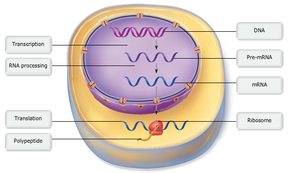






0 Response to "40 drag the labels onto the diagram to identify the parts of the cell."
Post a Comment