39 pupillary light reflex diagram
PDF REFLEX LAB - Advanced Overview 1 inch 3 inches Alphabet Concept Questions: Patellar Reflex and Pupillary Reflex Stations 1.In the following diagram, label as many as possible of the components involved in the patellar reflex: 2. In the following diagram, label the nervous system components involved in both the direct (same eye) and consensual (opposite eye) pupillary light reflex: PDF Anatomical pathway of pupillary light refles - Eye Learn ANATOMICAL PATHWAY OF PUPILLARY LIGHT REFLEX 1. Draw a labeled diagram of pupillary reflex pathway. (5) J2012 2. Pupillary pathways with diagram.(5) J2016 (1) Pupillary fibers in the nerve (2) Chiasmal decussation (3) Uncrossed fibers (4) Crossed fibers
Schematic diagram of the pupil light reflex showing ... A simple schematic diagram is shown in Fig. 1. The pupil area is determined by the interaction between constrict- ing and dilating mechanisms. Pupil contraction is caused by excitation of the...
Pupillary light reflex diagram
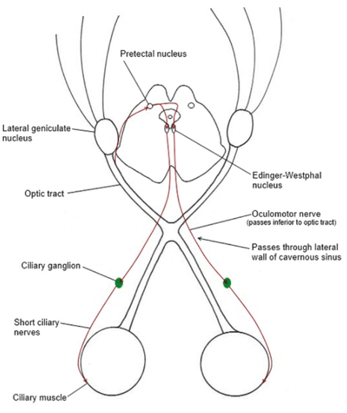
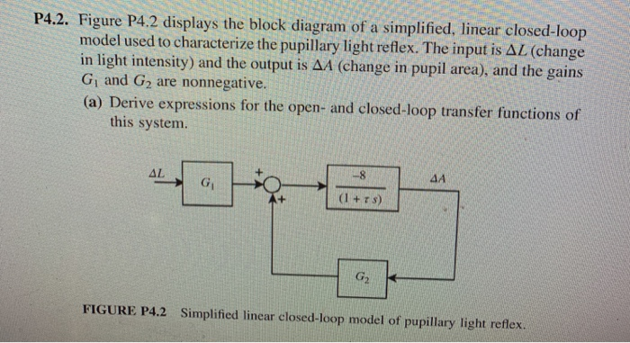
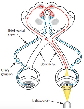
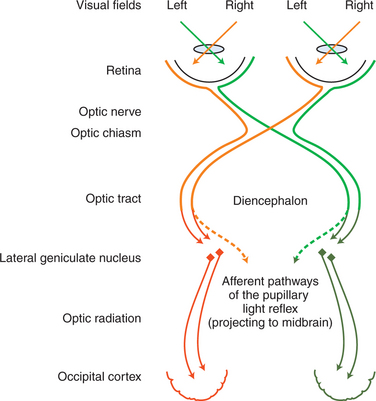
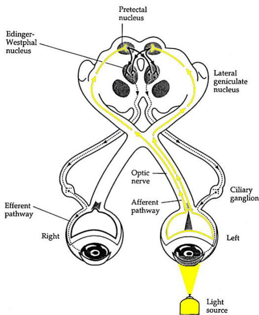
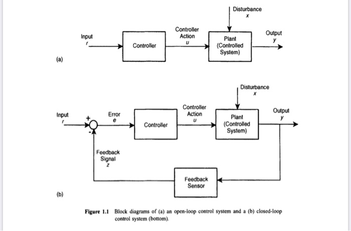
Pupillary Light Reflex - University of Georgia Dorsal view of direct pathway - This dorsal view of the afferent pathway shows how the impulse starts in the retina and crosses over in the optic chiasm to synapse in the pretectal nucleus of the thalamus. From here the pathway crosses back over to synapse in the nucleus of cranial nerve III. Solved P4.2. Figure P4.2 displays the block diagram of a ... Figure P4.2 displays the block diagram of a simplified, linear closed-loop model used to characterize the pupillary light reflex. The input is AL (change in light intensity) and the output is ΔΑ (change in pupil area), and the gains Gi and G2 are nonnegative. The Pupillary Light Reflex - YouTube Welcome to Soton Brain Hub - The Brain Explained!Dr James Booker officially joins our team and brings this really important clinical assessment to our catalo...
Pupillary light reflex diagram. Schematic drawing of the pupillary light reflex pathway ... Schematic drawing of the pupillary light reflex pathway. By way of the optic tract the afferent pathway (1) of the pupillary system projects to the dorsal midbrain. From the pretectal area, the... PDF VISUAL REFLEXES with loss of consciousness. What other ... (Aon Fig. 2 - Near Reflex Diagram) 1. Dilated right pupil and pupillary reflex absent on the right no matter which eye you shine light in. Present on left no matter which eye you shine it on. 2. Near response reflex absent on the right. 3. Ptosis on the right. 4. Lateral strabismus on the right. 5. Upward gaze paralysis on the right. B. Absent Pupillary Light Reflexes | Signs - MedSchool The pupillary light reflexes rely on a reflex pathway with the optic nerve as the sensory nerve, the oculomotor nerve as the motor nerve and the midbrain as the processing centre. How to Elicit Shine a light into each pupil. Solved Bio 150 Human Anatomy & Physiology DCCC (IV ... - Chegg Transcribed image text: Bio 150 Human Anatomy & Physiology DCCC (IV.B) - Demonstrate Monosynaptic Reflexes - On the diagram below, label the level of the spinal cord associated with each of the somatic reflex tests performed in the virtual lab. (IV.C) - Pupillary Light Reflex Arc- List the specific components of this reflex are below. You may need to use other sources such as your text or the ...
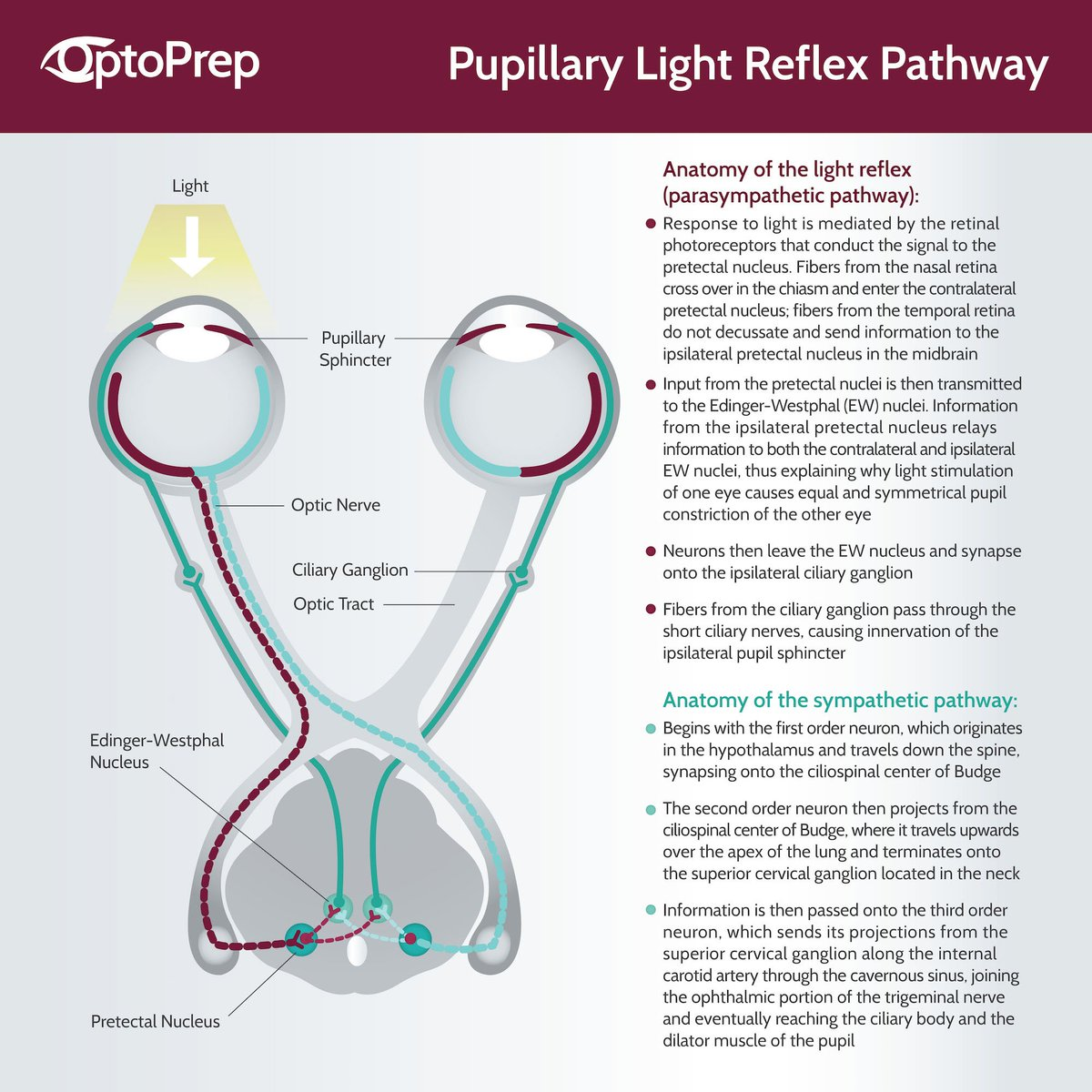
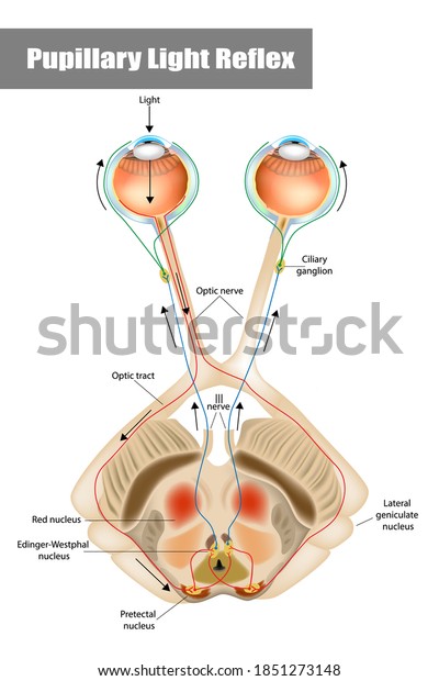
Pin on pupillary light reflex - Pinterest pupil light reflex pathway.red and blue lines represent the afferent pathway, the ganglion cell axons project to the pretectal region of the midbrain; green line represents the efferent pathway, the signal transmits from preganglionic parasympathetic fiber to ciliary ganglion, finally to the constrictor muscles through the short posterior ciliary … PDF Pupillary light reflex as an objective biomarker for early ... Pupillary Light Reflex (PLR) assessment The PLR-200™ (NeurOptics, Irvine, CA) monocular infrared pupillometer was used to quantify PLR under mesopic conditions (approximately 3 cd/m2). This is an FDA-approved hand-held cordless device that measures pupil size and dynamics (Figure 1). Reflexes 2 - Pupillary Light Reflex - YouTube - This tutorial is the second in a series of tutorials on the reflexes of the brainstem. This video covers the function a... Neuroanatomy of The Pupillary Light Reflex The diagram below shows the neuroanatomical pathways of the pupillary light The details of the pathway are detailed below the diagram. Afferent Pathway of Pupillary Light Reflex(solid yellow above): Light enters the pupiland stimulates the retina. Retinal ganglion cells transmit the light signal to the optic nerve
Sphincter pupillae: Origin, insertion, innervation,action ... Pupillary light reflex. The pupillary light reflex happens when the eyes are exposed to bright light and the amount of light that falls onto the retina needs to be decreased in order to maintain clear vision. The reflex arc includes optic nerve (CN II), pretectal nucleus of midbrain, accessory oculomotor nucleus and oculomotor nerve (CN III). Reflexes and the Eye - EyeWiki The pupillary light reflex is an autonomic reflex that constricts the pupil in response to light, thereby adjusting the amount of light that reaches the retina. Pupillary constriction occurs via innervation of the iris sphincter muscle, which is controlled by the parasympathetic system . Pupillary Light Reflex - StatPearls - NCBI Bookshelf Pupillary light reflexes are measured based on a 0 to 4+ gradient that considers the magnitude and speed of the light response. A normal, healthy adult patient is expected to have a 4+ response, which indicates a brisk, large response. Assessment of pupillary light reflex using a smartphone ... Pupil size . From the subjects of the current study (n=30), 60 paired comparisons were obtained. Images of pupillary light reflex acquired from the APP are presented in Fig. 3.Initial pupil size was 6.0±1.9 mm when measured using a PEN, and 5.8±1.8 mm when measured by the APP.
The assessment of pupils and pupillary reactions | Eye News
Reflexes | Boundless Anatomy and Physiology The stretch reflex (myotatic reflex) is a muscle contraction in response to stretching within the muscle. This reflex has the shortest latency of all spinal reflexes. It is a monosynaptic reflex that provides automatic regulation of skeletal muscle length. When a muscle lengthens, the muscle spindle is stretched and its nerve activity increases.

Medical Student Review: Pupillary light reflex- better than ...
Nonlinear Shunting Model of the Pupil Light Reflex The pupil light reflex is normally a feedback, closed-loop system regulating the retinal light flux. Many laboratory experiments use an open-loop configuration called the 'Maxwellian view' to avoid influences of the pupil size on the retinal light flux (Stark 1984). A light beam is brought to focus in the centre of the pupil.
Solved P4.2. Figure P4.2 displays the block diagram of a ...
The pupillary light reflex pathway | Neurology Objective: The anatomy of the human pupillary light reflex (PLR) pathway is a matter of debate. The aim of this study was twofold: namely, to investigate the association of a relative afferent pupillary defect (RAPD) in acquired suprageniculate lesions with the location and extent of the cerebral lesions. Further, we suggest a new strategy of lesion analysis by combining established techniques ...
Community Eye Health Journal » How to test for a relative ...
Pupillary Reflex - an overview | ScienceDirect Topics The pupillary light reflex (PLR) is the constriction of the pupil that is elicited by an increase in illumination of the retina. The direct PLR, present in virtually all vertebrates, is the constriction of the pupil in the same eye as that stimulated with light.

Schematic diagram of the pupil light reflex showing negative ...
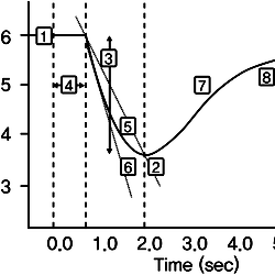
Pupillary light reflex - Wikipedia Referring to the neural pathway schematic diagram, the entire pupillary light reflex system can be visualized as having eight neural segments, numbered 1 through 8. Odd-numbered segments 1, 3, 5, and 7 are on the left. Even-numbered segments 2, 4, 6, and 8 are on the right.
Neuroophthalmology | Veterian Key
Pupillary Light & Blinking Reflexes Diagram - Quizlet Pupillary Light & Blinking Reflexes Diagram | Quizlet Science Medicine Ophthalmology Pupillary Light & Blinking Reflexes STUDY Learn Flashcards Write Spell Test PLAY Match Gravity + − THIS SET IS OFTEN IN FOLDERS WITH... Diencephalon 20 terms whitney_wallace1 PLUS Motor Sys 30 terms whitney_wallace1 PLUS Brainstem Nuc & tracts (CNV/VII/XI) 49 terms

JaypeeDigital | eBook Reader
Pupil Reflex: What Is The Function Of The Pupillary Light ... The pupillary light reflex is a reflex that controls the diameter of the pupil when it is exposed to varying intensities of light. This allows the eyes to adjust in response to bright or dim lights. Walk into any room and switch on the light; everything seems perfectly in its place. Now, switch off the light and try to see what is in the room.
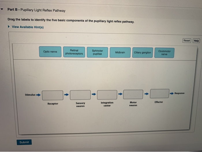
Solved Part B - Pupillary Light Reflex Pathway Drag the ...
A&P 2 Lab.docx - The Patella reflex 1. For the pathway ... The pupillary light reflex and consensual light reflex a. What is the response to shining the light in the right eye? the pupil will constrict b. Autonomic reflex c. For the stimulated right eye (pupillary light reflex): ipsilateral?

Pupillary light reflex pathway - Part 1 (Including Sample Questions)
Pupillary Light Reflex: study guides and answers on Quizlet Portable and easy to use, Pupillary Light Reflex study sets help you review the information and examples you need to succeed, in the time you have available. Use your time efficiently and maximize your retention of key facts and definitions with study sets created by other students studying Pupillary Light Reflex.

Eye movements, reflexes and control - ppt download
PDF VISUAL REFLEXES 4. Name three CNS and ... - University of Utah (Aon Fig. 2 - Near Reflex Diagram) 1. Dilated right pupil and pupillary reflex absent on the right no matter which eye you shine light in. Present on left no matter which eye you shine it on. 2. Near response reflex absent on the right. 3. Ptosis on the right. 4. Lateral strabismus on the right. 5. Upward gaze paralysis on the right. B.

Pupillary light reflex
Pupillary light reflex & pupillary light reflex test Pupillary light reflex. The pupillary light reflex is an autonomic reflex that constricts the pupil in response to light, thereby adjusting the amount of light that reaches the retina 1).Pupillary constriction occurs via innervation of the iris sphincter muscle, which is controlled by the parasympathetic system 2).. Testing of the pupillary light reflex is useful to identify a relative ...

Pathophysiology of Pupillary Reflexes -
The Pupillary Light Reflex - YouTube Welcome to Soton Brain Hub - The Brain Explained!Dr James Booker officially joins our team and brings this really important clinical assessment to our catalo...
PLOS ONE: Glutamatergic Neurotransmission from Melanopsin ...
Solved P4.2. Figure P4.2 displays the block diagram of a ... Figure P4.2 displays the block diagram of a simplified, linear closed-loop model used to characterize the pupillary light reflex. The input is AL (change in light intensity) and the output is ΔΑ (change in pupil area), and the gains Gi and G2 are nonnegative.
Pupillary Light Reflex | Arizona RETINA Project
Pupillary Light Reflex - University of Georgia Dorsal view of direct pathway - This dorsal view of the afferent pathway shows how the impulse starts in the retina and crosses over in the optic chiasm to synapse in the pretectal nucleus of the thalamus. From here the pathway crosses back over to synapse in the nucleus of cranial nerve III.

Stability analysis of reduced and original pupillary light ...
Pupil

Schematic drawing of the pupillary light reflex pathway. By ...

47 ideas de Pares craneales | pares craneales, neurología ...

9 Visual field defects ideas | visual, optic nerve, vision ...
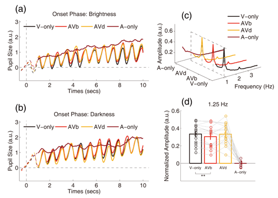
Pupillary Light Reflex Can Be Inhibited by Multisensory ...

Parasympathetic Pupillary Light Reflex Pathway - Neuroanatomy ...

Pupil light reflex pathway. Red and blue lines represent the ...

Pupillary light reflex... Afferent:2nd N... Efferent:3rd N ...
Brain Sciences | Free Full-Text | Using System Identification ...

Renewed Attention on the Pupil Light Reflex: Trends in ...
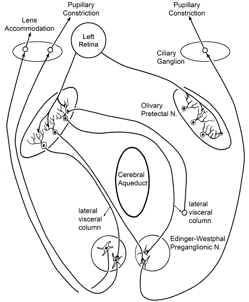
Figure 16 | Pupillary light reflex circuits in the macaque ...

King - pupillary light reflex (583) Diagram | Quizlet
Pupillary Light Reflex (PLR) oder Fotopupillary: Stock ...

File:PupillaryLightReflexNeuralPathway.jpg - Wikimedia Commons

Ophthalmology-Notes And Synopses - Pupillary light reflex Vs ...
![PDF] Eyeing up the Future of the Pupillary Light Reflex in ...](https://d3i71xaburhd42.cloudfront.net/88836be43482650dfdb4fc0cc47ca6e2d6afd759/7-Figure4-1.png)
PDF] Eyeing up the Future of the Pupillary Light Reflex in ...

Doctors Gates: Diagram shows pathway of the pupillary light ...

Pupillary Light Reflex (Labeled) on Meducation

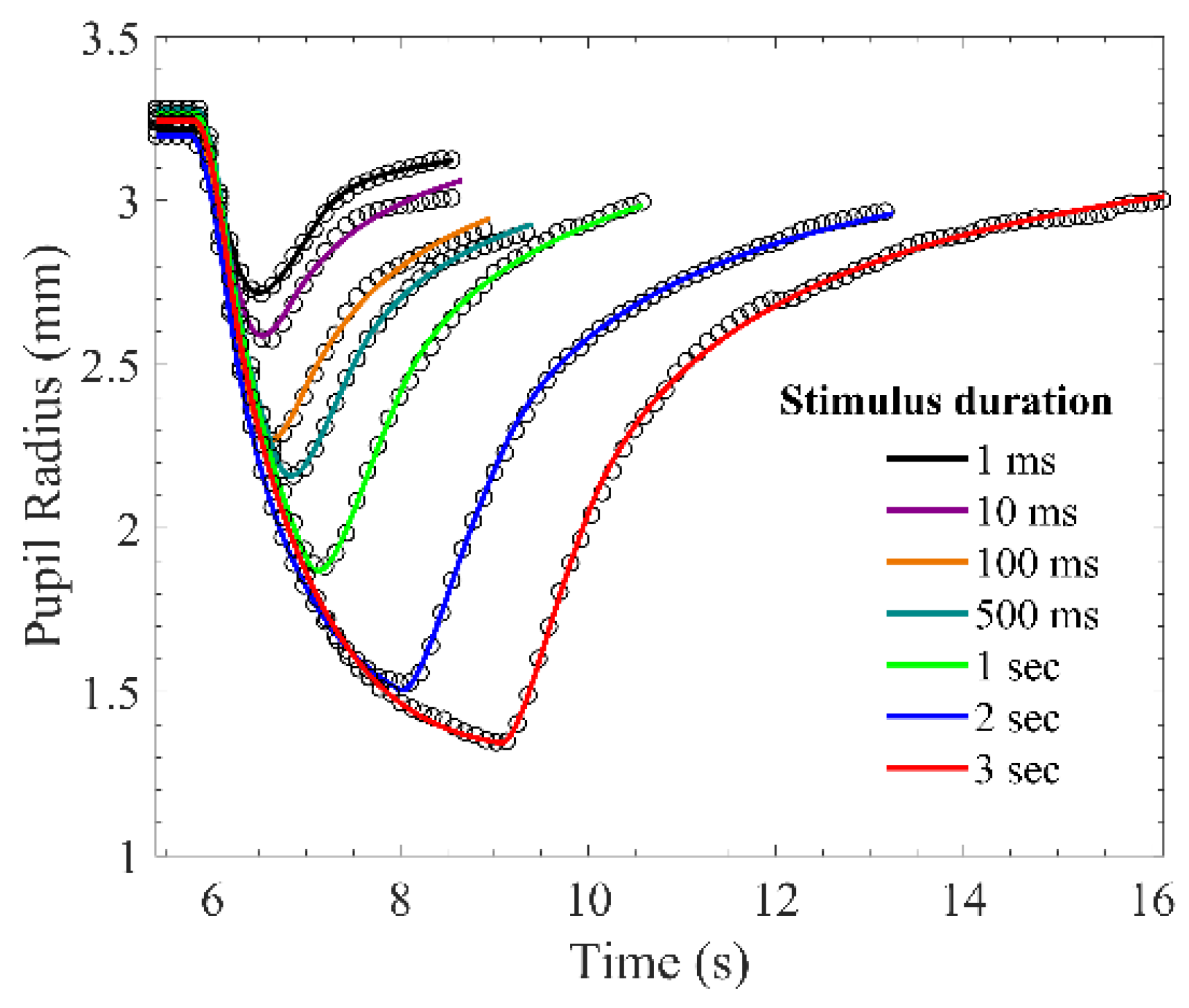
Mouse pupillary light reflex with 5 s of light stimulation ...
Solved P1.2. The pupillary light reflex is another classic ...

Pupillary reflexes physiology

The light reflex pathway showing the afferent path (red) and ...

B4W4 Visual Reflexes and Visual Field Defects-Tessema ...
Schematic diagram of the pupillary light response ...
0 Response to "39 pupillary light reflex diagram"
Post a Comment