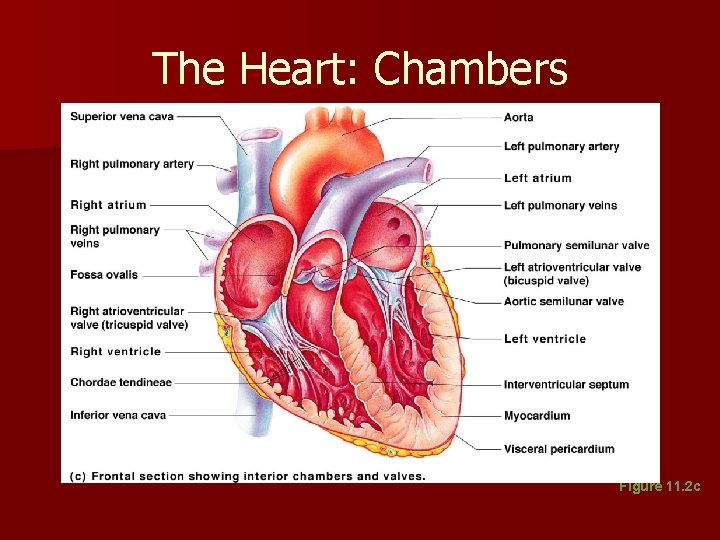
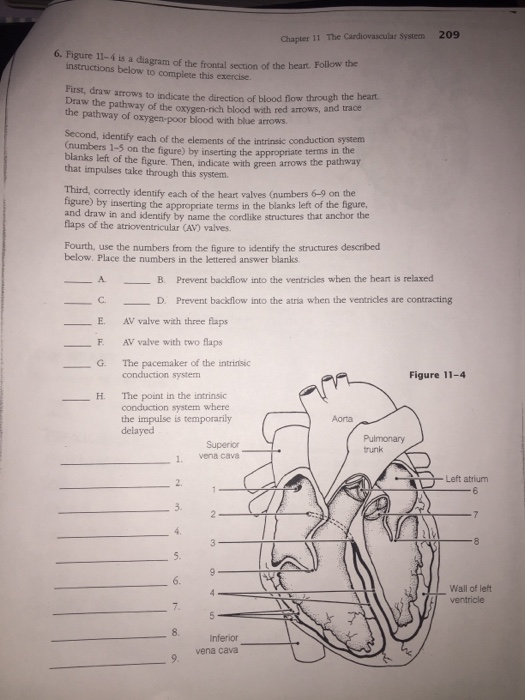
36 figure 11-4 is a diagram of the frontal section of the heart
Carefully study the diagram of the human respiratory system with labels A, B, C and D. Select the option which gives correct identification and main function and /or characteristic.(A) (i) Trachea: It is supported by bony rings for conducting inspired air.(B) (ii) Ribs: When we breathe out, ribs are A transverse section through the heart slightly above the level of the atrioventricular septum reveals all four heart valves along the same plane (Figure 11). The valves ensure unidirectional blood flow through the heart. Between the right atrium and the right ventricle is the right ...
The beating heart of the DX7 is a Hitachi 63B03 microprocessor, an enhanced version of Motorola's venerable late 70s 8-bit CPU built under license as a second-source supplier 4. Clocked at around 2MHz, it provides the synthesizer a steady pulse, albeit not the arithmetic power required for real-time digital synthesis 5.
Figure 11-4 is a diagram of the frontal section of the heart
Categories loading · When refering to evidence in academic writing, you should always try to reference the primary (original) source. That is usually the journal article where the information was first stated. In most cases Physiopedia articles are a secondary source and so should not be used ... The heart pumps around 5.7 litres of blood in a day throughout the body. The heart is situated at the centre of the chest and points slightly towards the left. On average, the heart beats about 100,000 times a day, i.e., around 3 billion beats in a lifetime. The average male heart weighs around 280 to 340 grams (10 to 12 ounces). The nasal cavity is a roughly cylindrical, midline airway passage that extends from the nasal ala anteriorly to the choana posteriorly.[1] It is divided in the midline by the nasal septum. On each side, it is flanked by the maxillary sinuses and roofed by the frontal, ethmoid, and sphenoid sinuses in an anterior to posterior fashion.[1] While seemingly simple, sinonasal anatomy is composed of ...
Figure 11-4 is a diagram of the frontal section of the heart. Your heart is an amazing organ. It continuously pumps oxygen and nutrient-rich blood throughout your body to sustain life. This fist-sized powerhouse beats (expands and contracts) 100,000 times ... The midsagittal section of the brain shows the three major parts of the brain, which are the cerebrum, cerebellum, and brainstem.These brain parts are marked with visible gross features like the gyri (singular: gyrus) and sulci (singular: sulcus) of the cerebrum. They are each also divided into subparts or regions for simplified localization of structures, for example, the brainstem is ... It is also known as Y-X plane or Frontal planes; the coronal plane divides the body into ventral (front) and dorsal (back) portions.This plane also gives a clear image of the posterior and anterior portions of the body. The coronal planes intersect the median plane at a 90-degree angle and show the anatomical body parts into front and back halves. Plan, Section, and Elevation are different types of drawings used by architects to graphically represent a building design and construction. A plan drawing is a drawing on a horizontal plane showing a view from above. An Elevation drawing is drawn on a vertical plane showing a vertical depiction. A section drawing is also a vertical depiction ...
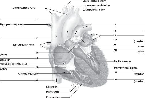
Anatomy and Physiology is a dynamic textbook for the yearlong Human Anatomy and Physiology course taught at most two- and four-year colleges and universities to students majoring in nursing and allied health. A&P is 29 chapters of pedagogically effective learning content, organized by body system, and written at an audience-appropriate level. Blood now returns to the heart from the lungs by way of the pulmonary veins (8) and goes into the left atrium (LA) (9). When the LA contracts, blood travels through the mitral valve (10) and into the left ventricle (LV) (11). The LV is a very important chamber that pumps blood through the aortic valve (12) and into the aorta (13). The aorta ... Below are more details regarding each section of the axial skeleton. Axial Skeleton: Skull The skull is located at the top portion of the human body and helps make up the head and face. Function and anatomy of the heart made easy using labeled diagrams of cardiac structures and blood flow through the atria, ventricles, valves, aorta, pulmonary arteries veins, superior inferior vena cava, and chambers. Includes an exercise, review worksheet, quiz, and model drawing of an anterior vi
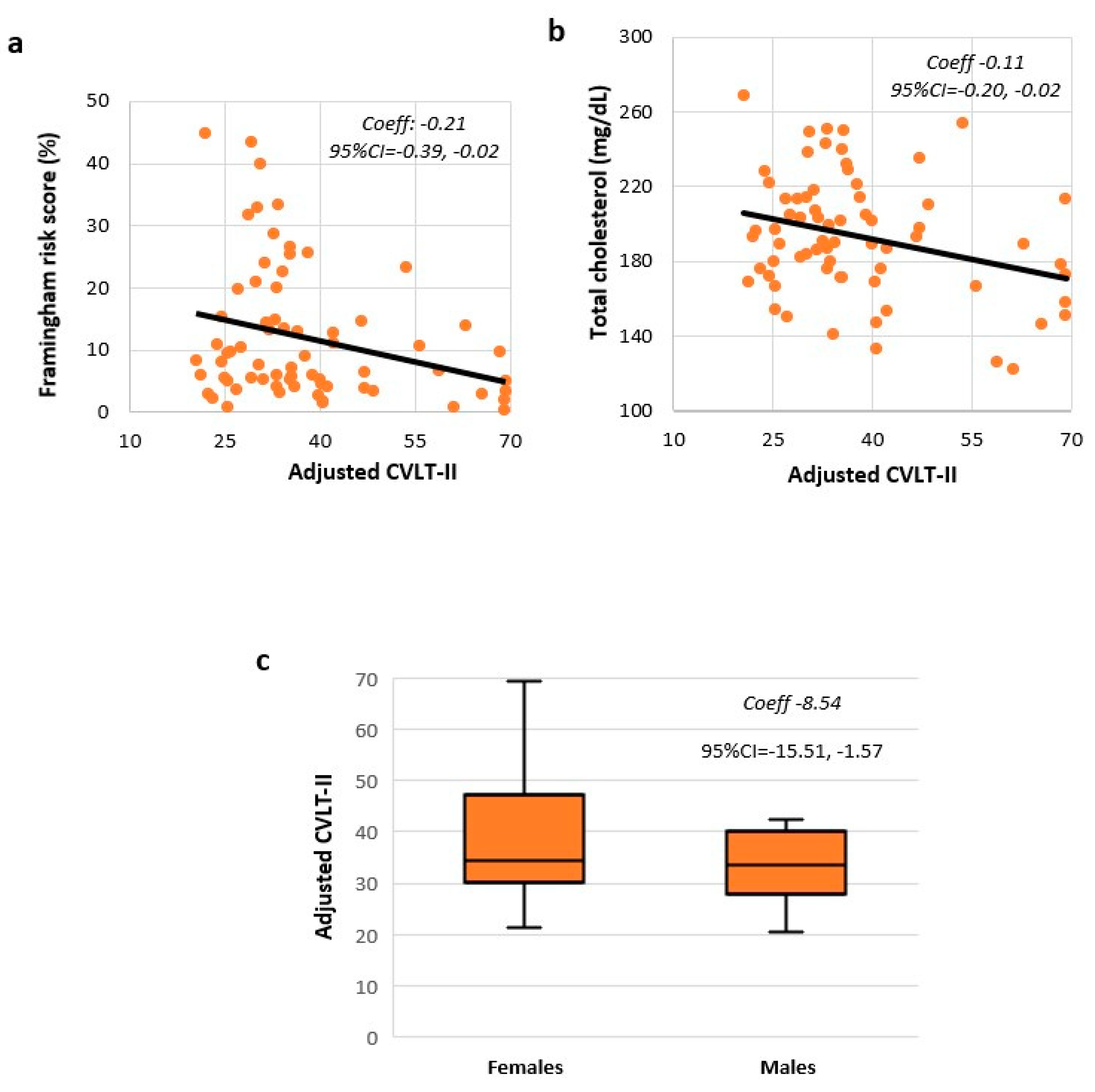
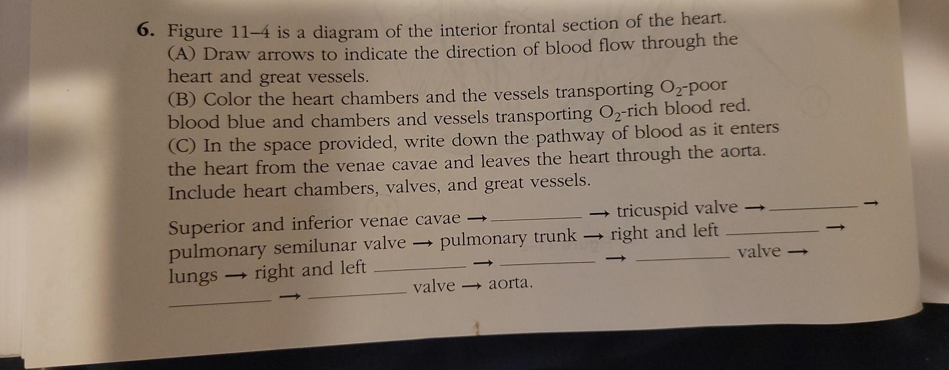
Features include 4: increased left ventricular internal end-diastolic diameter (LVIDd) parasternal long axis LVIDd >5.3 cm (females) or >5.9 cm (males) elevated left ventricular volumes. diastolic volumes >104 mL (females) or >155 mL (males) systolic volumes >49 mL (females) or >58 mL (males) The first major branch off of the aorta and the major artery to the forelimbs and head. Superior Vena Cava. receives blood from the head and arms and chest and empties into the right atrium of the heart. Ascending Aorta. the ascending part of the aorta as it emerges from the left ventricle. Figure 11-4 Is A Diagram Of The Interior Frontal Section Of The Heart. (A) Draw Arrows To Indicate The Direction Of Blood Flow Through The Heart And Great Vessels. (B) Color The Heart Chambers And The Vessels Transporting O2-poor Blood Blue And Chambers And Vessels Transporting O2-rich Blood Red. (C) In The Space Provided, Write Down The Pathway … Please note that this is just a preview of a ... Muscular System. The muscular system is responsible for the movement of the human body. Attached to the bones of the skeletal system are about 700 named muscles that make up roughly half of a person's body weight. Each of these muscles is a discrete organ constructed of skeletal muscle tissue, blood vessels, tendons, and nerves.
Images Pcmac Org
Stroke, a cerebrovascular accident, is prevalent across patient populations and can be a significant cause of morbidity and mortality. Stroke can be categorized as ischemic, hemorrhagic, or subarachnoid. Among ischemic strokes, the Trial Org 10172 in Acute Stroke Treatment (TOAST) classification is used to subdivide the categories that include cardioembolism, small-vessel occlusion, large ...

The Gut Microbiome In Neurological Disorders The Lancet Neurology
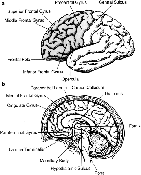
The frontal lobe, parietal lobe, occipital lobe, and temporal lobe have been associated with different functions ranging from reasoning to auditory perception. Frontal Lobe This lobe is located at the front of the brain and is associated with reasoning, motor skills, higher level cognition, and expressive language.

The Pathophysiology Of Cigarette Smoking And Cardiovascular Disease An Update Journal Of The American College Of Cardiology
The cerebral cortex has 4 main lobes - frontal lobe, parietal lobe, occipital lobe, and temporal lobe - and their location, function, and anatomy all differ. We will use labeled diagrams and lateral images of the brain (side views) to walk through each lobe of the cerebrum. Every EZmed post is filled with simple tricks to remember the content ...

Frontal Section Of The Heart Diagram Diagram Quizlet
Figure 11-4 is a diagram of the frontal section of the heart instructions below to complete this exercise Follow the First, draw arrows to indicate the direction Draw the pathway of the b the of blood flow through the heart. pathway of o t oxygen-rich blood with red arrows, and trace y xygen-poor ...

Uga Anatomy And Physiology 2 Lab Manual
6. Figure 11—4 is a diagram of the frontal section of the heart. Follow the instructions below to complete this exercise. First, draw arrows to indicate the direction of blood flow through the heart. Draw the pathway of the oxygen-rich blood with red arrows, and trace the pathway of oxygen-poor ...

17 Parts Of Your Body Ideas Body Circulatory System Human Brain Facts
Answer -. When it pumps the blood to the organs, the heart is contracted . When it receives the blood from the lungs, the heart is relaxed . In this figure, contraction in both ventricles is due to the fact that blood is pumping out. In the left ventricle, the oxygenated blood is pumping to other parts of the body.
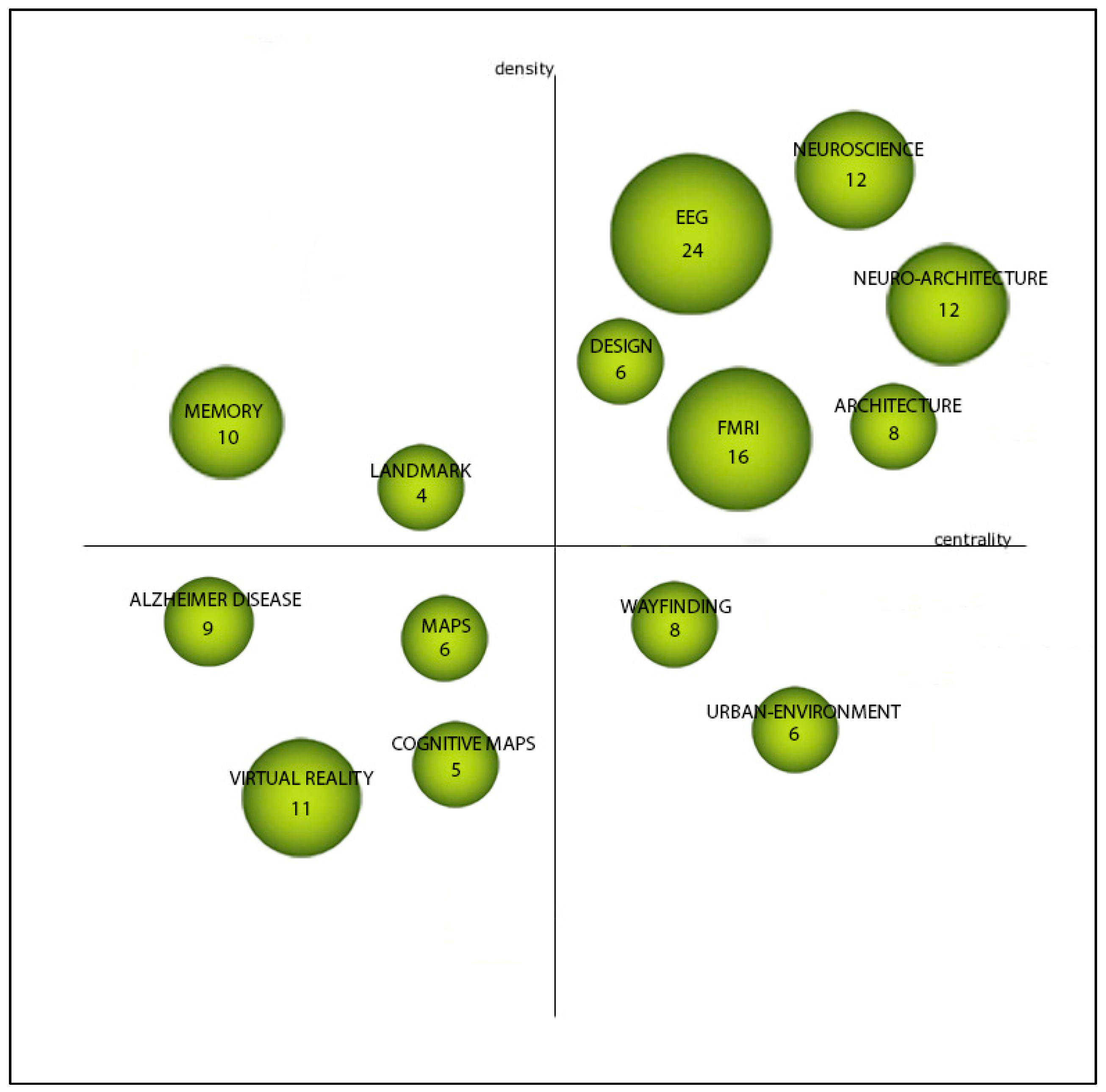
Ejihpe Free Full Text Neuroarchitecture Assessment An Overview And Bibliometric Analysis Html
Identify Various Parts Of A Human Heart: Trivia Quiz. The heart is the most important organ in the body. It is in charge of keeping the processes within the body moving by facilitating the transfer of blood throughout the body. The quiz below is to test out interesting facts you may know about the heart.

The Anatomy Of The Human Frontal Lobe Sciencedirect
The heart is upside down at this point, with the ventri-cle above the two incoming vessels. Around week 5, the heart curves back on itself in an “S” turn, creating the familiar anatomy, with the atria on top (Fig-ure 11.3). The adult heart is shown in . Figure 11.4.

Adventitial Elastolysis Is A Primary Event In Aneurysm Formation Journal Of Vascular Surgery
Four Chambers of the Heart and Blood Circulation. The shape of the human heart is like an upside-down pear, weighing between 7-15 ounces, and is little larger than the size of the fist. It is located between the lungs, in the middle of the chest, behind and slightly to the left of the breast bone. The heart, one of the most significant organs ...
Pathway
Cross section through the thalamus: Diagram Orienting yourself within such a cross section is easy. The star of the show (brain) is easily recognizable because it appears highly convoluted, full of ridges (gyri) and indentations (sulci).The paired thalami appear as two circular masses in the midline, forming the walls of the third ventricle.The neurocranium appears as a meshwork (trabecular ...
Cardiovascular System The Cardiovascular System N A Closed
The diagram in Figure 1 shows a section through the human heart, seen from the front. Figure 1 . Kingsmead Technology College Page 5 (a) Draw a ring around the correct answer to complete each sentence. (i) The wall of the heart is made mostly of epithelial glandular muscular tissue.
Chapter 44 Solutions Laboratory Manual For Human Anatomy Physiology Fetal Pig Version 2nd Edition Chegg Com
Besides, the data in Figure 1 shows that sometimes the amplitude data close to the radar is invalid and sometimes is large enough to saturate the receiver, especially in high sea states. To simplify this problem, in subsequent sections, we substitute the frontal 100 samples by the mean of the samples between positions 100 and 150.

Comparative Analysis Of The Combined Petrosal And The Pretemporal Transcavernous Anterior Petrosal Approach To The Petroclival Region In Journal Of Neurosurgery Ahead Of Print Journals
Figure 11-4 is a diagram of the frontal section of the heart. Follow the instructions below to complete this exercise. First, draw arrows to indicate the direction of blood flow through the heart. Draw the pathway of the oxygen-rich blood with red arrows, and trace the pathway of oxygen-poor ...
Brain Sciences Free Full Text A Retrospective Exploratory Analysis On Cardiovascular Risk And Cognitive Dysfunction In Multiple Sclerosis Html
A transverse section through the heart slightly above the level of the atrioventricular septum reveals all four heart valves along the same plane (Figure 19.1.11). The valves ensure unidirectional blood flow through the heart. Between the right atrium and the right ventricle is the right ...

Chapter 11 The Cardiovascular System Part I Ppt Video Online Download
209 Chapter 11 The Cardiovascular System 6. Figure 11-4 is a diagram of the frontal section of the heart instructions below to complete this exercise Follow the First, draw arrows to indicate the direction Draw the pathway of the b the of blood flow through the heart. pathway of o t oxygen-rich blood with red arrows, and trace y xygen-poor blood with blue arrows.
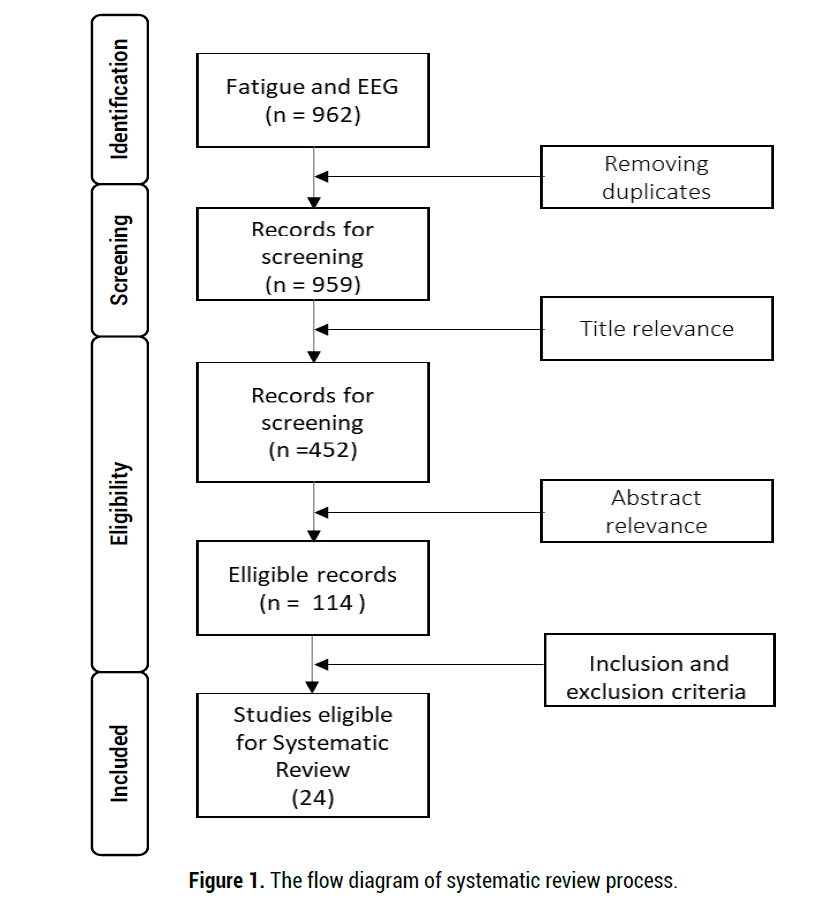
Systematic Review On The Use Of Electroencephalogram In Detecting Work Fatigue
Abnormal heart sounds are called , and usually indicate valve problems. 5. Figure 11-4 is a diagram of the frontal section of the heart. Follow the instructions below to complete this exercise. (a) Correctly identify each of the heart valves (numbers 6-9 on the figure) by inserting the appropriate terms in the blanks left of the figure.

Chapter 20 The Cardiovascular System The Heart Ppt Download
Abnormal heart sounds, or (10), usually indicate valve problems. 6. Figure 11—4 is a diagram of the frontal section of the heart. Follow the instructions below to complete this exercise. The colored arrows indicate the flow of oxygenated (red) and deoxygenated (blue) blood through the heart.

The Neurobiology Of Pain Perception In Normal And Persistent Pain Pain Management
Figure 11-4 is a diagram of the frontal section of the heart instructions below to complete this exercise Follow the First, draw arrows to indicate the direction Draw the pathway of the b the of blood flow through the heart. pathway of o t oxygen-rich blood with red arrows, and trace y xygen-poor ...

Ch 11 4 Brain Frontal Diagram Quizlet
An understanding of brain arterial vascular territories is important in understanding stroke and complications from surgery and endovascular procedures.. Although one could be excused for thinking that within the brain, such a carefully organized organ, blood supply would be constant, the truth is that a great deal of variety exists.
Solved 209 Chapter 11 The Cardiovascular System 6 Figure Chegg Com
One of the key steps in interpreting an electrocardiogram (EKG) is determining the electrical axis of the heart. Being able to determine the electrical axis can give insight into underlying disease states and help steer the differential diagnosis towards or away from certain diagnoses. Herein, we will discuss what makes up the electrical axis, ventricular (QRS) axis, axis classifications ...

The Neurobiology Of Pain Perception In Normal And Persistent Pain Pain Management
This is a plane that divides the body into superior and inferior portions. This plane runs perpendicular to the coronal and median planes and in an upright human is parallel, or horizontal, to the ...
Solved 6 Figure 11 4 Is A Diagram Of The Interior Frontal Chegg Com
Figure 11-4 is a diagram of the frontal section of the heart. Follow the instructions below to complete this exercise. (a) Correctly identify each of the heart valves (numbers 6-9 on the figure) by inserting the appropriate terms in the blanks left of the figure.

Cardiovascular Workbook Ola Docx The Cardiovascular System The Major Structures Of The Cardiovascular System The Heart And Blood Vessels Play A Vital Course Hero
The cardiovascular system circulates oxygen and nutrients throughout the body. PIXOLOGICSTUDIO/Science Photo Library/Getty Images Heart . The heart is the organ that supplies blood and oxygen to all parts of the body. This amazing muscle produces electrical impulses through a process called cardiac conduction.These impulses cause the heart to contract and then relax, producing what is known as ...
Web As Uky Edu
Coronal sections of the brain Author: Lorenzo Crumbie MBBS, BSc • Reviewer: Walter Muruet Last reviewed: October 25, 2021 Reading time: 13 minutes In clinical practice, the nervous system is usually visualised in sections that cut through one of the three main orthogonal planes: sagittal, coronal or horizontal .Each of these planes provides the clinician with information that allows the ...
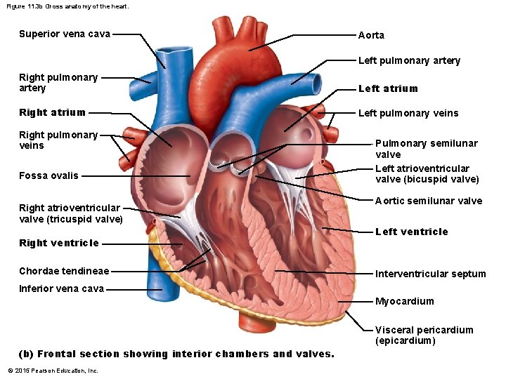
Figure 11 2 Heart Wall And Coverings 2015
Figure 11.4). It supplies oxygen- and nutrient-rich blood to all body organs. Because the left ventricle pumps blood over the much longer systemic path-way through the body, its walls are substantially thicker than those of the right ventricle (Figure 11.5), and it is a much more powerful pump.

Internal Features Of Heart Frontal Diagram Quizlet
The cerebral cortex (cortex of the brain) is the outer grey matter layer that completely covers the surface of the two cerebral hemispheres. It is about 2 to 4 mm thick and contains an aggregation of nerve cell bodies. This layer is thrown into complex folds, with elevations called gyri and grooves known as sulci. The cerebral cortex is quite distinct from the cerebrum (forebrain) which ...

Gross Anatomy Of The Heart Frontal Section Diagram Quizlet
Figure 11 4 is a diagram of the frontal section of the heart. 1 draw arrows to indicate the direction of blood flow through the heart. Around week 5 the heart curves back on itself in an s turn creating the familiar anatomy with the atria on top fig ure 113. Figure 11 4 is a diagram of the frontal section of the heart instructions below to complete this exercise follow the first draw arrows to ...

Internal Features Of The Heart Frontal Section Diagram Quizlet
300 Hillvue Lane, Pittsburgh, PA 15237 · Congratulations to all of our honor roll students

Chapter 27 Heart Anatomy Bio 140 Human Biology I Textbook Libguides At Hostos Community College Library
209 Chapter 11 The Cardiovascular System 6. Figure 11-4 is a diagram of the frontal section of the heart instructions below to complete this exercise Follow the First, draw arrows to indicate the direction Draw the pathway of the b the of blood flow...

The Heart Ppt Video Online Download
25. Figure 5—11 is a diagram of the articulated pelvis. Identify the bones and bone markings indicated by leader lines on the figure. Select different colors tor the structures listed below and use them to color the coding circles and corresponding structures in the figure.

Chapter 20 The Cardiovascular System The Heart Ppt Download
The nasal cavity is a roughly cylindrical, midline airway passage that extends from the nasal ala anteriorly to the choana posteriorly.[1] It is divided in the midline by the nasal septum. On each side, it is flanked by the maxillary sinuses and roofed by the frontal, ethmoid, and sphenoid sinuses in an anterior to posterior fashion.[1] While seemingly simple, sinonasal anatomy is composed of ...
Attention And The Frontal Cortex Springerlink
The heart pumps around 5.7 litres of blood in a day throughout the body. The heart is situated at the centre of the chest and points slightly towards the left. On average, the heart beats about 100,000 times a day, i.e., around 3 billion beats in a lifetime. The average male heart weighs around 280 to 340 grams (10 to 12 ounces).

Third Universal Definition Of Myocardial Infarction Circulation
Categories loading · When refering to evidence in academic writing, you should always try to reference the primary (original) source. That is usually the journal article where the information was first stated. In most cases Physiopedia articles are a secondary source and so should not be used ...
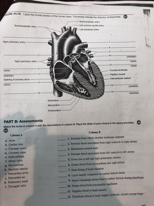
Solved Gure 44 15 Label This Frontal Section Of The Human Chegg Com

Figure 35 3 Frontal Section Of Human Heart Diagram Quizlet
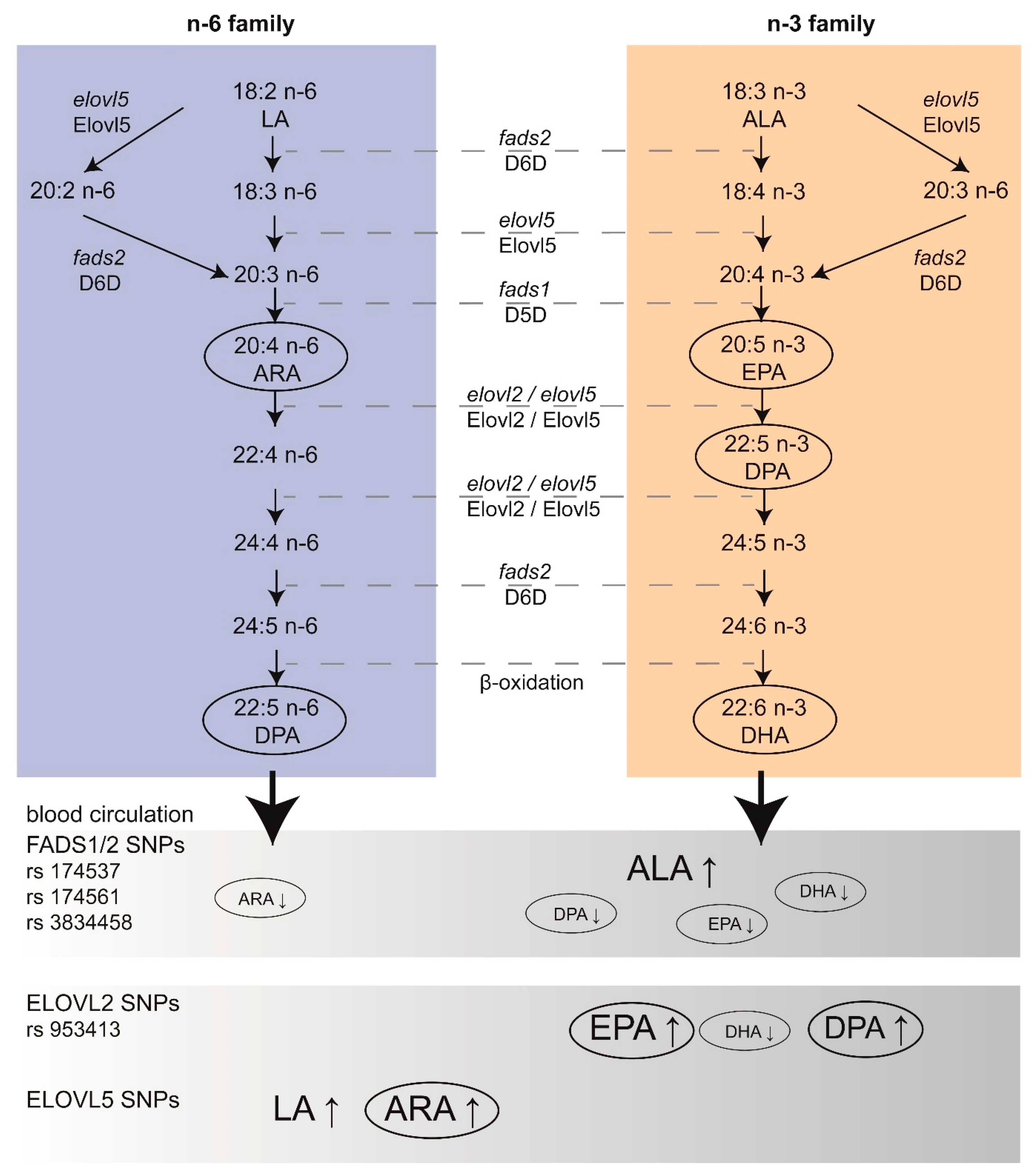
Nutrients Free Full Text Perinatal Dietary Polyunsaturated Fatty Acids In Brain Development Role In Neurodevelopmental Disorders Html
0 Response to "36 figure 11-4 is a diagram of the frontal section of the heart"
Post a Comment