37 diagram of compact bone
30.06.2021 · The ear canal, or auditory canal, is a tube that runs from the outer ear to the eardrum. The ear has outer, middle, and inner portions. The ear canal and outer cartilage of the ear make up the ... Drag the labels onto the diagram to identify the various types of synarthroses and amphiarthroses. 1. Suture 2. Gomphosis 3. Synchodrosis 4. Synostosis 5. Syndesmosis 6. Symphysis . Drag the labels to identify the structures within a synovial joint. 1. Medullary Cavity 2. Articular 3. Metaphysis 4. Spongy 5. Periosteum 6. Fibrous joint cap 7. Synovial 8. Joint Cavity 9. Compact bone. Drag and ...
05.04.2017 · Axial skeleton diagram. The appendicular skeleton consists of the pelvic girdle, the shoulder blades and arm bones and the legs and feet. This part of the endoskeleton protects and supports the limbs. Appendicular skeleton diagram. Movement. Bones, when supported by the function of muscles, deliver the capacity of locomotion (movement). The muscles are attached to the bone via …

Diagram of compact bone
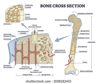
The walls of the diaphysis are composed of dense and hard compact bone. ... The top of this diagram shows the cross section of a generic bone with three ... Andrew Kirmayer A diagram of the anatomy of a bone, showing the compact bone. Human bone generally comprises osseous tissue, an outer coating called a periosteum, and bone marrow.The two main structural components typically include spongy bone on the interior, with an outer layer of compact bone. Usually found in long bones of the body, it consists of units called osteons, each of which is ... Bone Diagram Forehead (Frontal bone) Nose bones (Nasals) Cheek bone (Zygoma) Upper jaw (Maxilla) Lower jaw (Mandible) Breast bone (Sternum) Upper arm bone (Humerus) Lower arm bone (Ulna) Thigh bone (Femur) Collar bone (Clavicle) Toe bones (Phalanges) Ankle bones (Tarsals) Kneecap (Patella) Shin bone
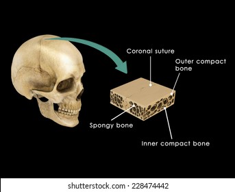
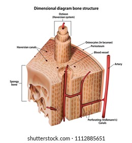
Diagram of compact bone. The compact bone is the main structure in the body for support, protection, and movement. Due to the strong nature of compact bone, compared to spongy bone, it is the preferred tissue for strength. Spongy bone is used for more active functions of the bones, including blood cell production and ion exchange. However, compact bones also serve a function in storing and releasing calcium to the ... Compact bone Although compact bone appears solid to the unaided eye, microscopically it contains considerable detail. The structural unit of compact bone is the osteon, or Haversian system (Fig. 6.5). Each osteon appears as a cylindrical unit consisting of 3–20 concentric lamellae of bone matrix surrounding the central osteonal canal Compact bone accounts for 80% of the bones in the human body. Compact bone, as opposed to spongy bone, is made of cylindrical units, called osteons, that are tightly formed together. As compact ... Compact bone forms the outer shell of the antler, and its greater density and stiffness provide strength for fighting. Spongy bone makes up about half the diameter of an average antler (McDonald et al. 2005), although there is considerable variation among animals. Antler Conformation. Antler shape or form, known as conformation, is highly variable and depends on age, genetics, and nutrition ...
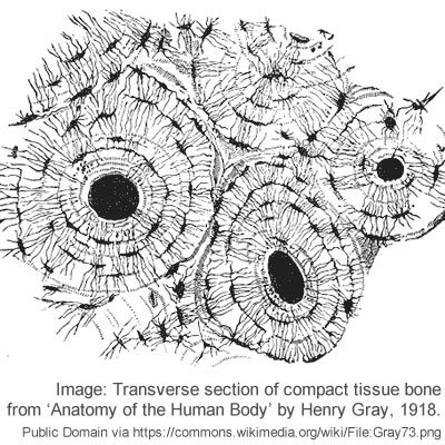
osteon diagram TS. The diagram above shows a transverse view of an osteon (Haversian system) - the basic unit of compact bone. diagram of haversian canal. May 13, 2019 - Compact Bone Diagram Long Bone Compact Bone And Spongy Bone Youtube. Compact Bone Diagram Osteon Compact Bone Ap Pinterest Anatomy Human ... Circuit diagram 150 KB Download Gendex DenOptix Dental X-Ray System Service manual ... Ziehm 7000, Compact, Vista C-Arm Maintenance procedure 750 KB Download prohibited by Ziehm. Support is not desired. Ziehm 7000 Plus C-Arm Service manual 6.3 MB Download prohibited by Ziehm. Support is not desired. Ziehm 8000 Service manual 9.0 MB Download prohibited by Ziehm. Support is not … Compact bone, dense bone in which the bony matrix is solidly filled with organic ground substance and inorganic salts, leaving only tiny spaces that contain ...
Compact bone is dense so that it can withstand compressive forces, while spongy bone (also called cancellous bone) has open spaces and is supportive, but also lightweight and can be readily remodeled to accommodate changing body needs. Compact Bone . Compact bone is the denser, stronger of the two types of osseous tissue (Figure 6.3.6). It makes up the outer cortex of all bones and is in ... Bone remodeling, the final phase of bone healing, goes on for several months. In remodeling, bone continues to form and becomes compact, returning to its original shape. In addition, blood circulation in the area improves. Once adequate bone healing has occurred, weightbearing (such as standing or walking) encourages bone remodeling. D. on cancellous, but not compact bone. E. in interstitial areas. on the surface of the bone. If an X-ray shows a black area in the region of the epiphyseal plate, A. the bone is fractured. B. the epiphyseal plate has not completely ossified. C. growth of the bone is complete. D. the cartilage is absent. E. marrow is forming in the cancellous bone. the epiphyseal plate has not completely ... A diagram of the anatomy of a bone, showing the compact bone. The cells of compact bone, which is also called cortical bone, appear to be tightly packed into a solid mass. Although the calls are close together, this type of bone is not completely solid. There are small canals that run through the bone, which allow blood vessels to penetrate it.

Diagram Of Compact Bone Open Educational Resource Oer Unsyiah Library
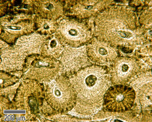
Figure 6.12 Diagram of Compact Bone (a) This cross-sectional view of compact bone shows the basic structural unit, the osteon. (b) In this micrograph of the osteon, you can clearly see the concentric lamellae and central canals. LM × 40.

Anatomy Microscopic Diagram Of Compact Bone Diagram Quizlet
A typical long bone shows the gross anatomical characteristics of bone. The structure of a long bone allows for the best visualization of all of the parts of a bone (Figure 1). A long bone has two parts: the diaphysis and the epiphysis. The diaphysis is the tubular shaft that runs between the proximal and distal ends of the bone.

Tampilan Petugas Diagram Of Spongy Bone
Compact bone is the denser, stronger of the two types of bone tissue ( [link] ). It can be found under the periosteum and in the diaphyses of long bones, where it provides support and protection. Diagram of Compact Bone. (a) This cross-sectional view of compact bone shows the basic structural unit, the osteon.

Bones Test Review Ppt Download
The fist is compact and thus effective as a weapon. It also provides protection for the fingers. It also provides protection for the fingers. [30] [31] [32] However, this is not widely accepted to be one of the primary selective pressures acting on hand morphology throughout human evolution, with tool use and production being thought to be far more influential.

Anatomy Ch 6 The Structure Of Compact Bone Diagram Quizlet
The pubic bone articulates with the ilium and the ischium on each hip. The internal surface forms the wall of the lesser pelvis and is the point of origin for a portion of the obturator internus muscle. The pubic bone is divisible into a body, a superior ramus, and an inferior ramus. The Body of the Pubic Bone. Body of pubic bone: (1) sacrum, (2) ilium, (3) ischium, (4) pubis: 4a-body; 4b ...

Compact Bone Diagram Quizlet
An equilibrium between osteoblasts and osteoclasts maintains bone tissue. Compact Bone. Compact bone consists of closely packed osteons or haversian systems.

Bone Cells And Structure Anatomy And Physiology
Bone Diagram Forehead (Frontal bone) Nose bones (Nasals) Cheek bone (Zygoma) Upper jaw (Maxilla) Lower jaw (Mandible) Breast bone (Sternum) Upper arm bone (Humerus) Lower arm bone (Ulna) Thigh bone (Femur) Collar bone (Clavicle) Toe bones (Phalanges) Ankle bones (Tarsals) Kneecap (Patella) Shin bone

This Picture Shows Different Views Of Compact Bone Important Labels To Look At Here Are Lamellae Ce Nursing School Survival Health Education Nursing Programs
Andrew Kirmayer A diagram of the anatomy of a bone, showing the compact bone. Human bone generally comprises osseous tissue, an outer coating called a periosteum, and bone marrow.The two main structural components typically include spongy bone on the interior, with an outer layer of compact bone. Usually found in long bones of the body, it consists of units called osteons, each of which is ...

Bone Histology General Overview Compact Bone Is The Dense Part Of Download Scientific Diagram
The walls of the diaphysis are composed of dense and hard compact bone. ... The top of this diagram shows the cross section of a generic bone with three ...
Compact Bone Tissue Images Stock Photos Vectors Shutterstock

Structure Of Compact Bone Quiz

Structure Of Bones Biology For Majors Ii

Compact Bone 1 Diagram Quizlet

Anatomy Of Compact Bone Compact Bone Tissue Osteon Diagram Stock Illustration Illustration Of Cells Exchanging 191577368
What Is The Structure And Function Of The Compact Bone Socratic

Microscopic Labeling Of Compact Bone Diagram Quizlet
Compact Bone Tissue Images Stock Photos Vectors Shutterstock
Cartilage Bone Ossification The Histology Guide

Compact Bone Histology Diagram Quizlet

Homework Read Chap 6 Study All The Bone

Anatomy And Physiology Bones And Skeletal Tissue Anatomy Histology Anatomy Of The Ey Cell Diagram Skeletal System Anatomy Bones Human Anatomy And Physiology

Major Parts Of A Compact Bone Diagram Quizlet

Structure Of Compact Bone Longitudinal And Cross Sectional View Of Download Scientific Diagram

Long Bone Compact Bone And Spongy Bone Anatomy Bones Human Anatomy Drawing Human Anatomy

Microscopic Anatomy Of Compact Bone Diagram Quizlet
2

Drawing Illustrates The Labelled Structures Of Compact And Spongy Bone Download Scientific Diagram

Bones Structure And Types Youtube
1

Compact Bone Diagram Koibana Info Anatomy Models Labeled Human Anatomy Model Anatomy Bones

Compact Bones Vs Spongy Bones What Is The Difference Diffzi
Compact Bone Tissue Images Stock Photos Vectors Shutterstock

Compact Bone Tissue Images Stock Photos Vectors Shutterstock

2cm Section Of Diaphysis Compact Bone Removed Download Scientific Diagram
Cartilage Bone Ossification The Histology Guide

Pin By Amy Schrader On A P Human Anatomy And Physiology Anatomy Bones Skeletal System Anatomy

6 Osteocytes Within Compact Bone Download Scientific Diagram
0 Response to "37 diagram of compact bone"
Post a Comment