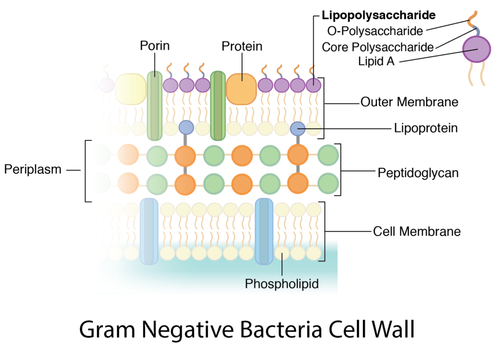
36 gram negative cell wall diagram
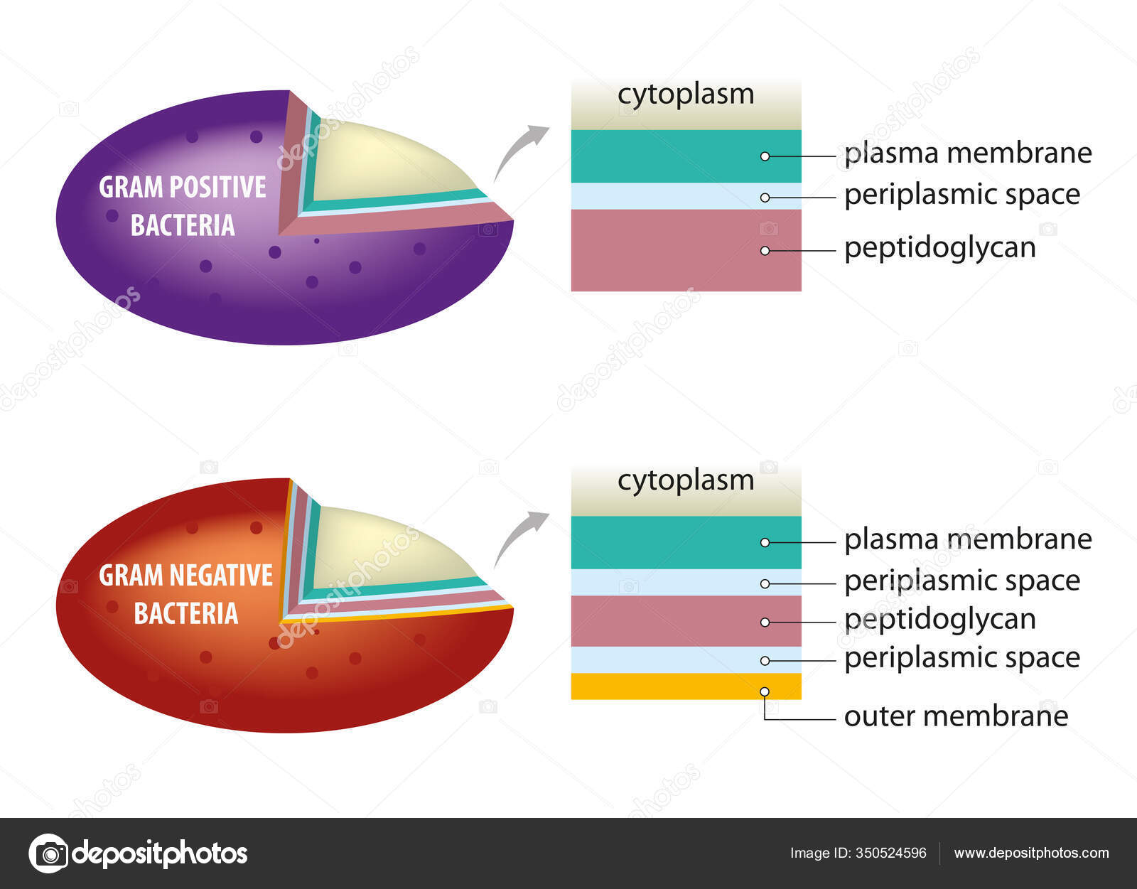
Periplasmic Space: This cellular compartment is found only in those bacteria that have both an outer membrane and plasma membrane (e.g. Gram negative bacteria).In the space are enzymes and other proteins that help digest and move nutrients into the cell. Cell Wall: Composed of peptidoglycan (polysaccharides + protein), the cell wall maintains the overall shape of a bacterial cell. 3.4 Bacterial Cell Walls 1. Describe peptidoglycan structure. 2. Compare and contrast the cell walls of typical Gram-positive and Gram-negative bacteria. 3. Relate bacterial cell wall structure to the Gram-staining reaction. 37
28.4.2017 · Cell Wall Definition. A cell wall is an outer layer surrounding certain cells that is outside of the cell membrane.All cells have cell membranes, but generally only plants, fungi, algae, most bacteria, and archaea have cells with cell walls.The cell wall provides strength and structural support to the cell, and can control to some extent what types and concentrations of molecules enter and ...

Gram negative cell wall diagram
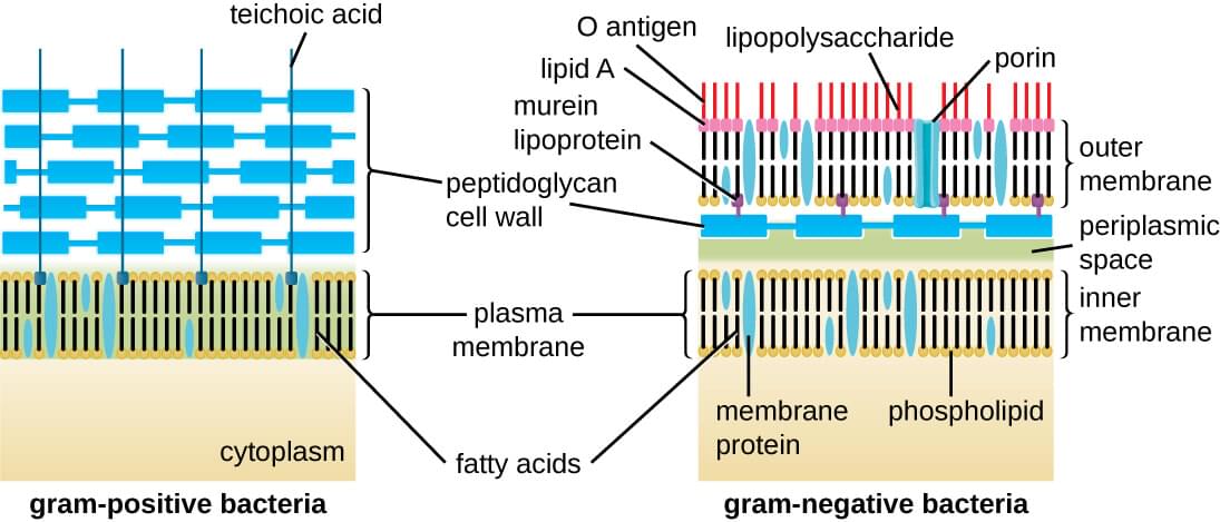
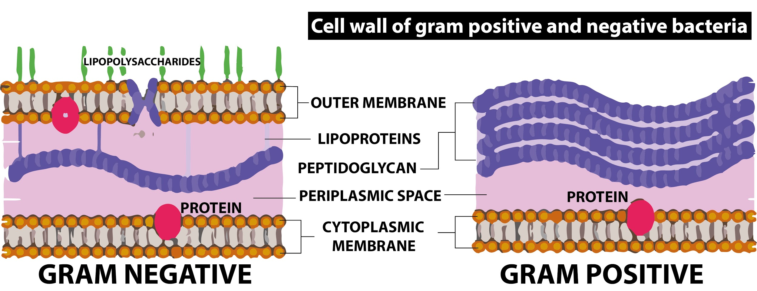
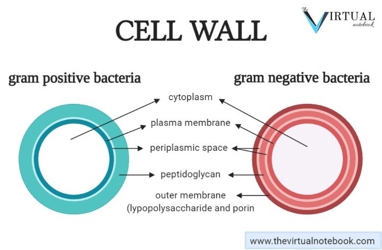
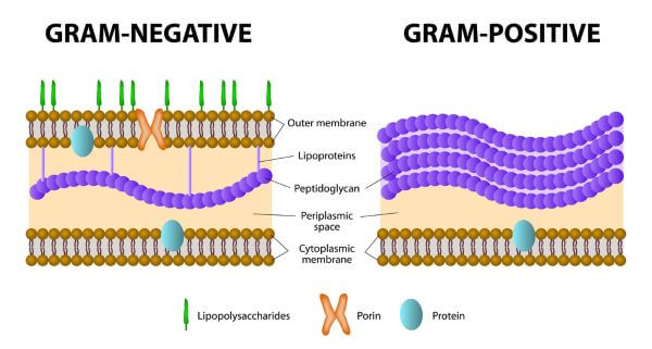
Download scientific diagram | Schematic structure of Gram-positive and Gram-negative cell walls. Gram-positive cell walls contain only one lipid plasma membrane and a thick peptidoglycan layer ... The cell wall of gram-negative bacteria is complex having a thin layer of the peptidoglycan layer of 2-7nm and a thick outer membrane of 7-8nm thick. Microscopically, there is a space that is seen between the cell membrane and the cell wall, known as the periplasmic space made up of periplasm. However it is found in both Gram-negative and Gram ... Periplasmic space occurs between plasma membrane and cell wall. Cell wall protects the bacterial cells against bursting in hypotonic solution. Wall is 20-80 nm thick in Gram positive bacteria. It is single layered and smooth. In Gram negative bacteria, wall is 8-12 nm thick, complex, wavy and two layered. The outer layer is also called outer ...
Gram negative cell wall diagram. 26.8.2019 · A cell wall is a rigid, semi-permeable protective layer in some cell types. This outer covering is positioned next to the cell membrane (plasma membrane) in most plant cells, fungi, bacteria, algae, and some archaea. Animal cells however, do not have a cell wall. The cell wall has many important functions in a cell including protection, structure, and support. In electron micrographs, the Gram-positive cell wall appears as a broad, dense wall 20-80 nm thick and consisting of numerous interconnecting layers of peptidoglycan (see Figs. 1A and 1B). Chemically, 60 to 90% of the Gram-positive cell wall is peptidoglycan. In Gram-positive bacteria it is thought that the peptidoglycan is laid down in cables ... The walls of gram-positive bacteria have simpler chemical structures compared to gram-negative bacteria. Gram-positive cell wall. Gram-positive cell wall is thick measuring about 15-80 nm and more homogenous compared to gram-negative cell wall. This cell wall consists of large amount of peptidoglycan arranged in several layers. Peptidoglycan in ... 21.8.2019 · Gram negative bacteria appear a pale reddish color when observed under a light microscope following Gram staining. This is because the structure of their cell wall is unable to retain the crystal violet stain so are colored only by the safranin counterstain. Examples of Gram negative bacteria include enterococci, salmonella species and ...
Start studying Gram negative cell wall. Learn vocabulary, terms, and more with flashcards, games, and other study tools. In addition, they may contain polysaccharide molecules. The gram negative cell wall contains three components that lie outside the peptidoglycan layer: 1. Lipoprotein. 2. Outer membrane and . 3. Lipopolysaccharide. Thus the different results in the gram stain are due to differences in the structure and composition of the cell wall. File:Gram negative cell wall.svg. Size of this PNG preview of this SVG file: 800 × 401 pixels. Other resolutions: 320 × 160 pixels | 640 × 320 pixels | 1,024 × 513 pixels | 1,280 × 641 pixels | 2,560 × 1,282 pixels | 1,486 × 744 pixels. A cell wall is a structural layer surrounding some types of cells, just outside the cell membrane.It can be tough, flexible, and sometimes rigid. It provides the cell with both structural support and protection, and also acts as a filtering mechanism. Cell walls are absent in animals but are present in most other eukaryotes including algae, fungi and plants and in most prokaryotes (except ...
Nov 13, 2021 · Gram-positive wall; Gram-negative wall; The gram-positive bacteria are classified so because of their thick cell wall that contains many layers of peptidoglycan and teichoic acid. Gram-negative bacteria, on the other hand, have a wall that is thin with a few layers of peptidoglycan that is surrounded by a second lipid membrane. This additional ... 4 Bacteria: Cell Walls . It is important to note that not all bacteria have a cell wall.Having said that though, it is also important to note that most bacteria (about 90%) have a cell wall and they typically have one of two types: a gram positive cell wall or a gram negative cell wall.. The two different cell wall types can be identified in the lab by a differential stain known as the Gram stain. Start studying Gram-Negative Bacterial Cell Wall, Unit 1. Learn vocabulary, terms, and more with flashcards, games, and other study tools. Jan 03, 2021 · The Gram-negative cell wall is composed of a thin, inner layer of peptidoglycan and an outer membrane consisting of molecules of phospholipids, lipopolysaccharides (LPS), lipoproteins and sutface proteins. The lipopolysaccharide consists of lipid A and O polysaccharide. The peptidoglycan portion of the Gram-negative cell wall is generally 2-3 ...
Download scientific diagram | Diagram demonstrating of the cell wall structure of (a) Grampositive bacterium, (b) Gram-negative bacterium, and (c) mycobacterium. from publication: Differentiation ...
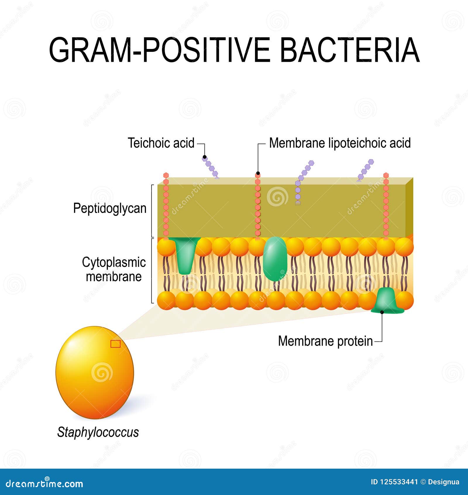
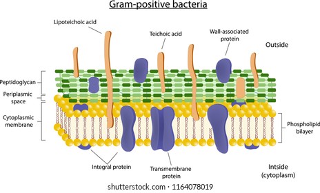
Differences between Gram-positive and Gram-negative bacteria cell wall The gram-Positive Cell wall of Bacteria. Bacterial cell wall that is gram-positive contains peptidoglycan and teichoic acids with some species having additional carbohydrates and proteins. The murein component is what gives shape to the gram-positive bacterial cell wall; it ...
In Figure 4.3, which diagram of a cell wall is a gram-negative cell wall? b. Each of the following statements concerning the gram-positive cell wall is true EXCEPT. it protects the cell in a hypertonic environment. The number of organelles such as chloroplasts, mitochondria, and rough endoplasmic reticulum is the same in all eukaryotic cells. ...
Gram-negative cell walls are strong enough to withstand ∼3 atm of turgor pressure (), tough enough to endure extreme temperatures and pHs (e.g., Thiobacillus ferrooxidans grows at a pH of ≈1.5) and elastic enough to be capable of expanding several times their normal surface area ().Strong, tough, and elastic … the gram-negative cell wall is a remarkable structure which protects the ...
14.10.2021 · Gram-negative bacteria, which do have an outer membrane, don't pick up the stain. For example, ... Delving beneath the cell wall and membrane, …
A: Gram Positive Bacteria Cell Wall B: Similarities C: Gram Negative Bacteria Cell Wall A B C Teichoic acid is present in the peptidoglycan layer P… View the full answer Transcribed image text : Complete this Venn diagram comparing Gram Positive and Gram Negative Cell Walls Venn Diagram
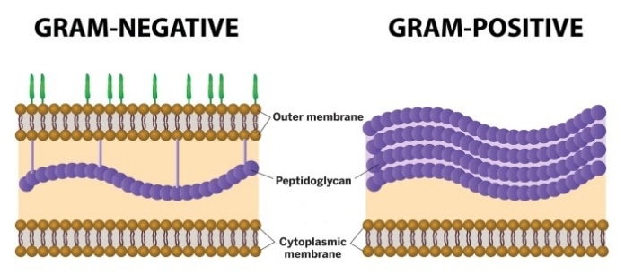
The key difference between gram positive and gram negative cell wall is that the gram positive cell wall has a thick peptidoglycan layer with teichoic acids while gram negative cell wall has a thin peptidoglycan layer surrounded by an outer membrane. Another major difference between gram positive and gram negative cell wall is that the gram positive cell wall stains in purple colour in grams ...
Gram-negative bacteria have a smaller amount of this rigid structure than do gram-positive bacteria. Periplasmic space. an open area between the cell wall and cell membrane in the cell envelopes of bacteria. gram negative bacteria have a more extensive space than do a gram positive bacteria. Cell membrane.
Gram staining Procedure. Retains the crystal violet dye and appear purple in colour. Does not retain the crystal violet dye and appear pink in colour. These differences between the Cell Wall of Gram-positive and Gram-negative Bacteria are classified based on their structure, composition of the cell and by the procedure of Gram staining technique.
In Gram-positive bacteria, peptidoglycan makes up as much as 90% of the thick cell wall enclosing the plasma membrane. Gram Staining! During Gram staining, these thick, multiple layers (20-80 nm) of peptidoglycan retain the dark purple primary stain crystal violet, whereas Gram-negative bacteria stain pink.
Gram-negative, Gram-positive, anaerobes: Rarely: aplastic anemia. Inhibits bacterial protein synthesis by binding to the 50S subunit of the ribosome Fosfomycin: Monurol, Monuril: Acute cystitis in women: This antibiotic is not recommended for children and 75 and up of age: Inactivates enolpyruvyl transferase, thereby blocking cell wall ...
Periplasmic space occurs between plasma membrane and cell wall. Cell wall protects the bacterial cells against bursting in hypotonic solution. Wall is 20-80 nm thick in Gram positive bacteria. It is single layered and smooth. In Gram negative bacteria, wall is 8-12 nm thick, complex, wavy and two layered. The outer layer is also called outer ...
The cell wall of gram-negative bacteria is complex having a thin layer of the peptidoglycan layer of 2-7nm and a thick outer membrane of 7-8nm thick. Microscopically, there is a space that is seen between the cell membrane and the cell wall, known as the periplasmic space made up of periplasm. However it is found in both Gram-negative and Gram ...
Download scientific diagram | Schematic structure of Gram-positive and Gram-negative cell walls. Gram-positive cell walls contain only one lipid plasma membrane and a thick peptidoglycan layer ...









0 Response to "36 gram negative cell wall diagram"
Post a Comment