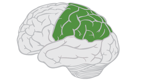
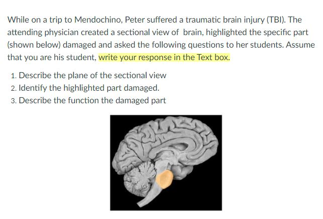
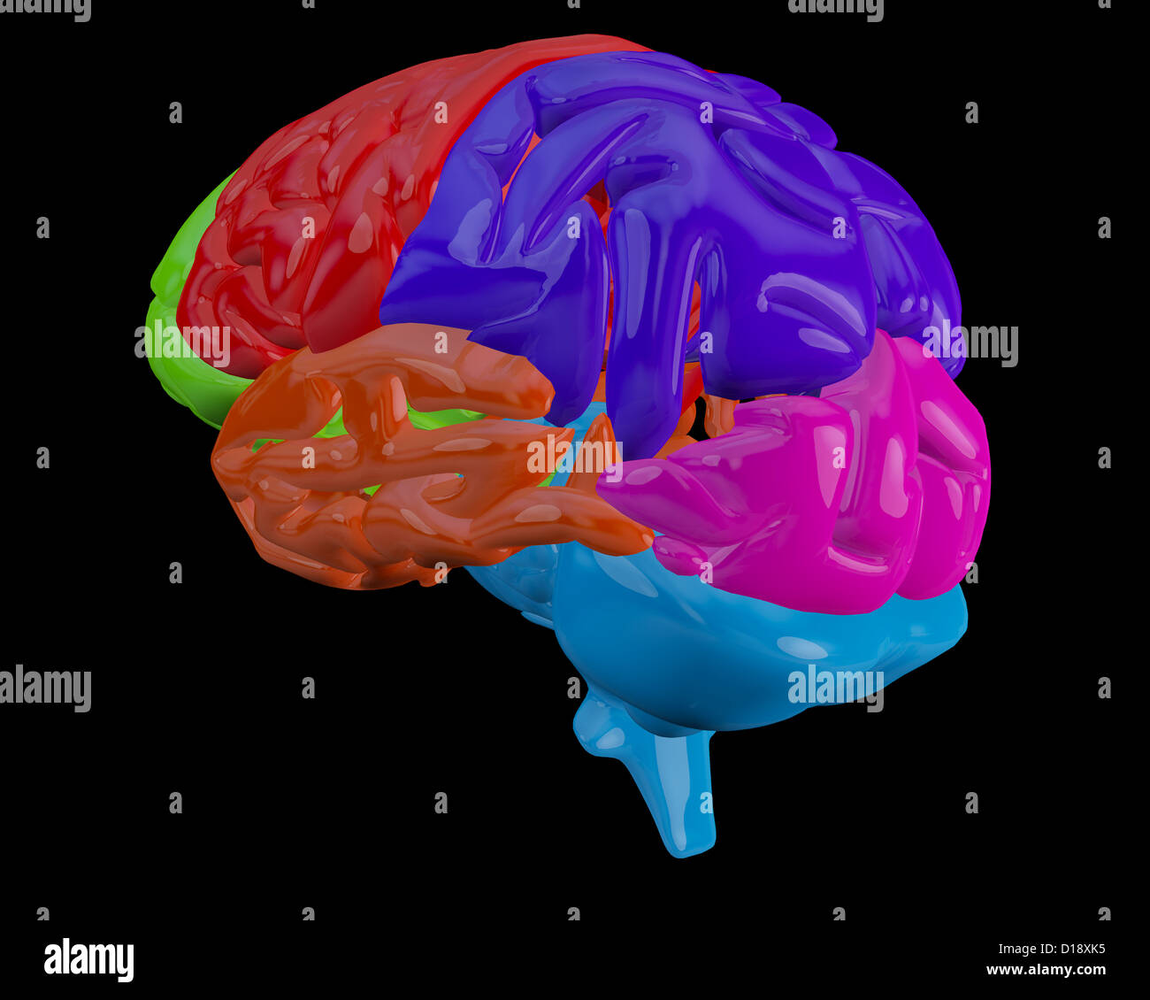
38 what part of the brain is highlighted in the diagram below?
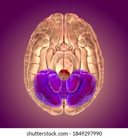
The highlighted part you are talking about is certainly the cerebellum. it is located in the posterior cranial fossa, below and at the back of the two hemispheres, it looks like a little brain. Its most important roles consist in the handling of the equilibrium of the body and learning ability.
retina converts light into electrical impulses that are sent to the brain through the optic nerve. Vitreous gel: The vitreous gel is a transparent, colorless mass that fills the rear ... parts of the eye, eye diagram, vitreous gel, iris, cornea, pupil, lens, optic nerve, macula, retina ...
human brain, if available), identify the areas and structures of the cerebral hemispheres described above. Then continue using the model and preserved brain along with the figures as you read about other structures. Diencephalon The diencephalon is embryologically part of the fore-brain, along with the cerebral hemispheres. See Figure 17.1 ...

What part of the brain is highlighted in the diagram below?
brain activity for teens in the area that processes motivation and pleasure than that used for decision making. This indicates that teens may focus more on rewards and less on risks when making decisions.) 2. Describe how each brain-imaging tool highlighted in the article teaches something different about the relationship between the brain
PLAY. What are the responsibilities of the region of the brain highlighted below? Coordinating movement and balance by using information from sensory nerves, including hand-eye coordination. Which of the following structures or systems is correctly paired with its function? Nice work!
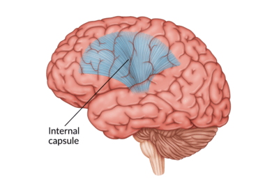
The cerebrum, the foremost part of the brain, is the largest part of the brain in humans comprising about 83% of total brain mass 8. It separates the left and right cerebral hemispheres from one another. 9. Gyri 10. The transverse fissure Question 2 0 / 0 pts The Brain 11.
What part of the brain is highlighted in the diagram below?.
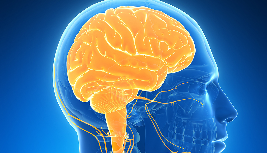
The brain is an organ that's made up of a large mass of nerve tissue that's protected within the skull. It plays a role in just about every major body system. The cerebrum is the largest part ...
Give the LETTER and NAME of the part of the brain responsible for: 1.4 The diagram below represents a human brain. 1.4.1 Memorising a cellular phone number (2) 1.4.2 Coordinating all voluntary movements (2) 1.4.3 Secreting hormones (2) 1.4.4 Connecting the two hemispheres of part B (2)
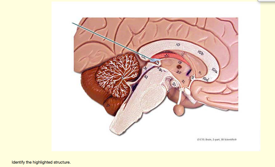
cerebellum Identify the highlighted structure. pons Identify the highlighted structure. cerebellum This part of the brain is responsible for coordination of fine motor skills. pons This part of the brain serves as a bridge between cerebellar hemispheres. (allowing one to juggle objects between two hands easily) corpora quadrigemini
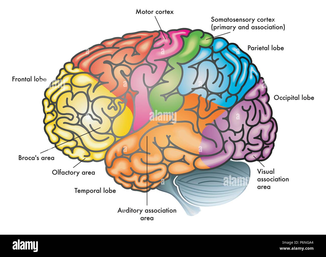
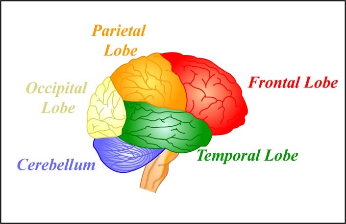
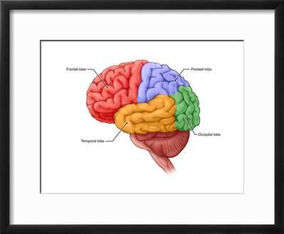
The brain is a 3-pound organ that contains more than 100 billion neurons and many specialized areas. There are 3 main parts of the brain include the cerebrum, cerebellum, and brain stem. The Cerebrum can also be divided into 4 lobes: frontal lobes, parietal lobes, temporal lobes, and occipital lobes.
D)transmit impulses from the effector to the brain 42.The diagram below represents a reflex arc. The function of the neuron labeled X is to A)an effector B)a motor neuron C)an interneuron D)a receptor 43.A reflex arc is illustrated in the diagram below. Structure X represents A)an antibody B)a pigment C)a neurotransmitter D)an antigen
QUESTION 33 The diagram below shows DNA of a hypothetical fruit fly gene ken (called ken and barbie, for my favorite gene). Several elements are highlighted by numbers 1-4. In wildtype fly larvae, the gene product (protein) is expressed in cells that will eventually become excitatory neurons. This gene is also expressed in the adult
the highlighted part you are talking about is certainly the cerebellum. it is located in the posterior cranial fossa, below and at the back of the two hemispheres, it looks like a little brain. Its most important roles consist in the handling of the equilibrium of the body and learning ability. Advertisement Answer 1.3 /5 5 merridyx2 Answer:
Label the following structures on the diagram below: frontal lobe, parietal lobe, occipital lobe, temporal lobe, cerebellum, brain stem, central sulcus, lateral sulcus, transverse fissure, precentral gyrus, postcentral gyrus Indicate (roughly), where Broca's area and Wernicke's area can be found. 2. What is the function of the corpus callosum? 3.
The height of the human brain is about 3.6 inches and it weighs about 4 to 5 lbs at birth and 3 lbs in adults. The total surface area of the cerebral cortex is about 2,500 cm2 and when stretched, it will cover the area of a night table. The brain is composed of 77 to 78% water and 10 to 12% lipids. It contains 8% proteins 1% carbohydrates, 2% ...
The diagram of the brain is useful for both Class 10 and 12. It is one among the few topics having the highest weightage of marks and is frequently asked in the examinations. A well-labelled diagram of a human brain is given below for further reference.
The diagram below shows a human brain. What part of the brain is labeled 3 in the diagram? A. frontal lobe B. temporal lobe C. occipital lobe D. parietal lobe Correct Answer: D. 15.
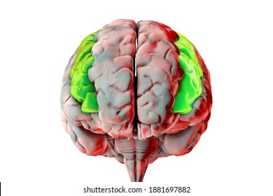
12 Dec 2018 — the part that is highlighted is the parietal lobe. musashixjubeio0 and 7 more users found this answer helpful. Thanks 4.2 answers · Top answer: it is the parietal lobe
Labeled brain diagram. First up, have a look at the labeled brain structures on the image below. Try to memorize the name and location of each structure, then proceed to test yourself with the blank brain diagram provided below. Labeled diagram showing the main parts of the brain.
The brain is one of the largest and most complex organs in the human body. It is made up of more than 100 billion nerves that communicate in trillions of connections called synapses. • The ...
The word 'cerebellum' literally means little brain. It is the second largest part of the brain, and is located at the back, below the occipital lobe, beneath the cerebrum and behind the brain stem. It contains an outer gray cortex and an inner white medulla, and has horizontal furrows, which makes it look different from the rest of the brain.
BI 335 - Advanced Human Anatomy and Physiology Western Oregon University Figure 4: Mid-sagittal section of brain showing diencephalon (includes corpus callosum, fornix, and anterior commissure) Marieb & Hoehn (Human Anatomy and Physiology, 9th ed.) - Figure 12.10 Exercise 2: Utilize the model of the human brain to locate the following structures / landmarks for the
What part of the brain is highlighted in the diagram below? (4 points) Frontal lobe Occipital lobe Temporal… Get the answers you need, now! kyliebruner kyliebruner 04/27/2021 Biology High School answered 3. What part of the brain is highlighted in the diagram below? (4 points) Frontal lobe Occipital lobe Temporal lobe Parietal lobe 1 See ...
about the whole brain that is pictured on the right. Use the drop-down text menu of parts of the brain to see each brain region. Brain structures are highlighted on the G2C Brain as you study each structure, and parts are labeled when "View Labels" is clicked. You can rotate the brain model to see the inferior (bottom), superior (top),
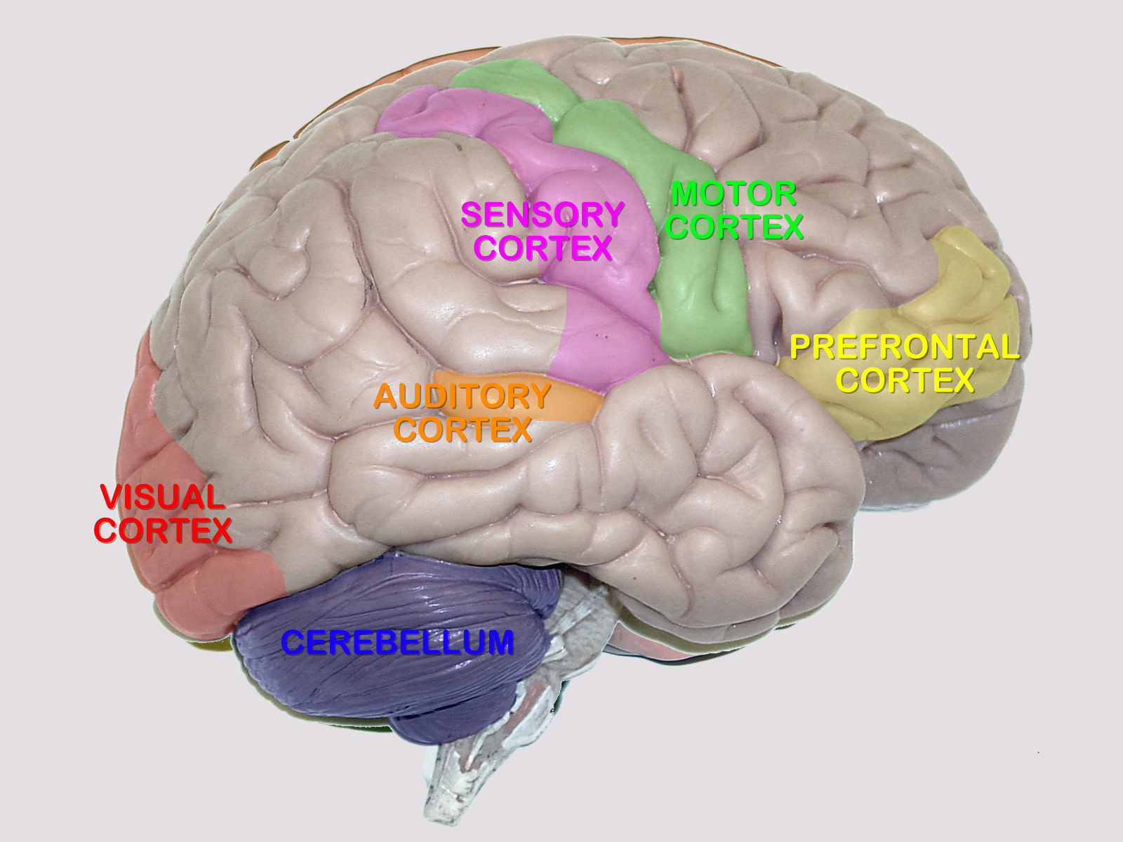
Cerebellum - the part of the brain below the back of the cerebrum. It regulates balance, posture, movement, and muscle coordination. Corpus Callosum - a large bundle of nerve fibers that connect the left and right cerebral hemispheres. In the lateral section, it looks a bit like a "C" on its side. Frontal Lobe of the Cerebrum
What part of the brain is highlighted in the diagram below? Image is highlighting the section at the rear of the brain, found at the back of each hemisphere ...1 answer · Top answer: The highlighted part you are talking about is certainly the cerebellum. it is located in the posterior cranial fossa, below and at the back of ...
John Medina · 2018 · Educationtheir ever-present molecular garbage, adding to brain health by getting rid of ... they share basic structures, highlighted in the simplified diagram below.
Tim Sole, Rod Marshall · 2009 · Games & Activities... routes home from the shopping mall , the shopper must live somewhere on the highlighted section or at the circled point in the first diagram below .
At a high level, the brain can be divided into the cerebrum, brainstem and cerebellum. Cerebrum The cerebrum (front of brain) comprises gray matter (the cerebral cortex) and white matter at its center. The largest part of the brain, the cerebrum initiates and coordinates movement and regulates temperature.
7 Sept 2021 — Just below the midbrain is the pons, and below the pons is the medulla. The medulla is the part of the brain stem closest to the spinal cord.
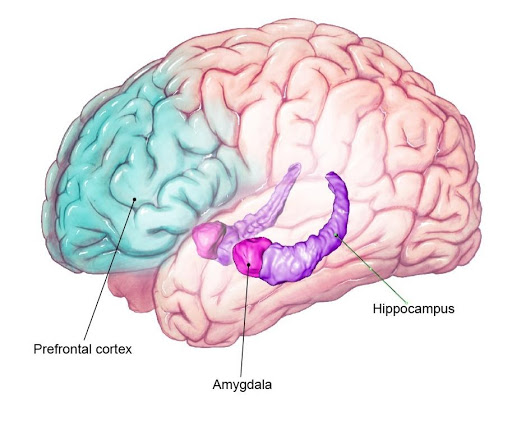
William J. Ray · 2019 · PsychologyBack to Figure Illustration of brain is shown in the diagram. The parts highlighted and labeled include Prefrontal cortex, Amygdala, and Hippocampus.
Back to The Brain and Learning. Support for LabX programming is generously provided by the Marian E. Koshland Endowment Fund
Nick & Bethan Redshaw... confirm that particular areas within the brain highlighted by the earlier ... of function in the brain and hemispheric lateralisation The diagram below ...
Brain diagram highlighting various parts of the human brain. ... The hypothalamus is a small and essential part of the brain, located precisely below the thalamus. It is considered the primary region of the brain, as it is involved in the following functions: Receives impulses;
Download scientific diagram | 1 A human brain with the cerebellum highlighted in purple. (Figure presented with permission, courtesy of the National Institute of Health, NIH) from publication ...









/auditory-cortex-highlighted-in-brain-149320441-58580ae05f9b586e028cf183.jpg)





/https://tf-cmsv2-smithsonianmag-media.s3.amazonaws.com/filer/Memory-hippocampus-brain-631.jpg)

0 Response to "38 what part of the brain is highlighted in the diagram below?"
Post a Comment