39 cat muscles labeled diagram
Cat dissection lab_labeled_images. 1. Cat Dissection Muscular Labs. 2. External oblique Pectroalis minor Pectoralis major Gastrocnemius Sartorius Tibialis anterior Gracilis. 3. Latissimus dorsi Levator scapula Lumbodorsal fascia Gluteal muscles Deltoid Biceps femoris Gastrocnemius Trapezius Sartorius Trapezius Semitendinosis External oblique ... Anatomy & Physiology Cat Muscles ... The deepest lateral muscles of the cat shoulder, brachium, and forearm. Supraspinatus Infraspinatus Teres major deltoideus Triceps brachii long head Triceps brachii lateral head Brachialis . Sartorius Sartoñus (cut)
Gain a comprehensive understanding of a cat's health with our veterinary guide to cat anatomy complete with diagrams, videos and simple explanations - cardiovascular, digestive, musckuloskeletal, respiratory, urogenital systems.

Cat muscles labeled diagram
Find the rectus femoris by separating the sartorius and biceps femoris. The rectus femoris is a deep muscle between the two. Locate the palmaris longus and the brachioradialis in the upper limb of the cat. Human Muscles Labeled Diagram For Kids Infographics Muscle. C Ous Atae M Figure 311 Illustrates The Ven Cheggcom. Royalty Free Stock Illustration Of Diagram Dorsal Ventral Cavities. A Motor And A Brake Two Leg Extensor Muscles Acting At The Same. Muscle Diagrams To Label Diagram Dorsal Cat Oasissolutionsco. Cat References Day 1 Terminology & External/Internal Anatomy. Pictures from iBook; Cat Skeleton Tutorial-Kenyon College; Day 2&3 Muscular System. Muscle Tables; Cutaneous, Shoulder & Back Muscles
Cat muscles labeled diagram. 27/05/2012 · The muscles of the lower extremity that can reach the TLF include the gluteus maximus and biceps femoris. Finally, the torso muscles involved in influencing this fascial and aponeurotic composite include the lumbar epaxial muscles and hypaxial muscles such as the TrA and the internal oblique, and possibly a small part of the external oblique. The following links will allow you to access real photographs of the cat muscular system. The purpose of these pages is to quiz your knowledge on the structures of the muscular system. Please try to answer all structures (or guess) before you look at the answers! Choose one of the following categories: Neck Muscles. Neck (Superficial) Structure. Palatoglossus arises from the palatine aponeurosis of the soft palate, where it is continuous with the muscle of the opposite side, and passing downward, forward, and lateralward in front of the palatine tonsil, is inserted into the side of the tongue, some of its fibers spreading over the dorsum, and others passing deeply into the substance of the organ to intermingle with … labeled-diagram-of-digestive-system. Labeled Diagram Of Digestive System Diagram - Labeled Diagram Of Digestive System Chart - Human anatomy diagrams and charts explained. This diagram depicts Labeled Diagram Of Digestive System with parts and labels.
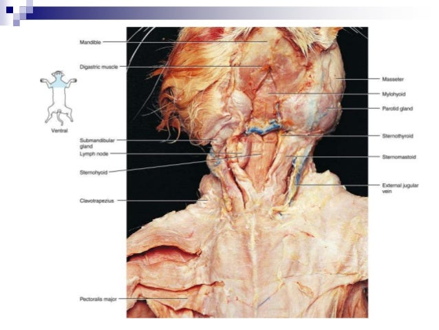
Cross-sectional labeled anatomy of the head and neck of the domestic cat on CT imaging (bones of the skull, cervical spine, mandible, hyoid bone, muscles of the neck, nasal cavity and paranasal sinuses, oral cavity, larynx) This is an online quiz called Cat Muscle Anatomy. From the quiz author. name the muscels in a cats back Your Skills & Rank. Total Points. 0. Get started! Today's Rank--0. Today 's Points. One of us! Game Points. 14. You need to get 100% to score the 14 points available. Actions. Add to favorites 7 favs. Add to Playlist. Questions. 20. Tries. Unlimited. Last Played. 15 Jan, 2022. Sound On/Off. From the quiz author. Muscles of a cats hind legs for BIOL-2401. Unit 21: Anatomy of the Domestic Cat 1. Muscles of the cat abdomen 1 2. Muscles of the cat abdomen 2 3. Muscles of the cat abdomen 3 4. Superficial muscles of the cat chest 5. Muscles of the back and shoulder of the cat 6. Muscles of the lateral thigh of the cat 7. Muscles of the medial thigh of the cat 1 ...
The cat's third eyelid is known as the nictitating membrane. It is located in the inner corner of the eye, which is also covered by conjunctiva. In healthy cats, the conjunctiva of the eyelids is not readily visible and has a pale, pink color. Integumental. The two main integumentary muscles of a cat are the platysma and the cutaneous maximus. 1. Given alongside is a diagram of a smear of human blood. Study the same and answer the questions that follow: (a) Name the parts 1, 2, 3 and 4 indicated by guidelines. Solution:-1 is Red Blood Cell (RBC) 2 is White Blood Cell (WBC) 3 is Blood Platelet. 4 is Blood Plasma. (b) Mention two structural differences between the parts labeled 1 and 2 ... Cat neck muscles anatomy The diagram alongside shows and arrangement of three pulleys A, B and C. The load is marked as L and the effort as E. (a) Name the pulleys A, B and C. (b) Mark in the diagram the direction of load (L), effort (E) and tension T 1 and T 2 in the two strings. (c) How are the magnitudes of L and E related to the tension T 1?
Diagram Cat Anatomy Muscles - Diagram Media
Cat muscles are one of the essential parts of feline anatomy. Muscles—skeletal, cardiac and smooth—are tissues that contract to allow for movement and force. Muscles can make a cat travel from one place to another or make organs function properly. Skeletal Muscles, Cardiac Muscles and Smooth Muscles ...
Shegüy, With you I learned what it is to love without possessing, to accompany without invading and to live without depending. Photography by: me (Shewylab).
The latest Lifestyle | Daily Life news, tips, opinion and advice from The Sydney Morning Herald covering life and relationships, beauty, fashion, health & wellbeing

Adductor Longus + Adductor Femoris + Semimembranosus ...
The vulva (plural: vulvas or vulvae; derived from Latin for wrapper or covering) consists of the external female sex organs.The vulva includes the mons pubis (or mons veneris), labia majora, labia minora, clitoris, vestibular bulbs, vulval vestibule, urinary meatus, the vaginal opening, hymen, and Bartholin's and Skene's vestibular glands.The urinary meatus is also included as it …
Cat dissection lab_labeled_images
The muscles of your body quiz trivia. The muscles of the body come in handy when it comes to controlling the movement of parts of the body. The muscles are divided into smooth, skeletal and cardiac muscles. Every body part has...

Muscles , 5 Cat Muscle Anatomy Diagram : Cat Muscles ...
Browse 500 sets of quiz cat muscles flashcards. Study sets Diagrams Classes Users. 31 Terms. tallmadgiraffe. Cat Muscles. pectoralis major. pectoralis minor. rectus abdominis. external oblique.
Startled blue-eyed cat
To play this quiz, please finish editing it. INSTRUCTOR-LED SESSION. Start a live quiz. SUPER. Classic. Students progress at their own pace and you see a leaderboard and live results. Instructor-paced BETA. Control the pace so everyone advances through each question together.

Superficial muscles of a cat : Biological Science Picture ...
Leg muscles are located closer to the body, allowing ostriches to run ... The life cycle of a frog is illustrated in the diagram. Fertilized eggs Embryos Adult frog ... Turtle Horse Wolf Tiger House cat Hair Sharp teeth Retractable claws Ability to purr Four limbs
Pretty boy
Human body muscle diagrams. Muscle diagrams are a great way to get an overview of all of the muscles within a body region. Studying these is an ideal first step before moving onto the more advanced practices of muscle labeling and quizzes. If you're looking for a speedy way to learn muscle anatomy, look no further than our anatomy crash courses .

Cat Muscles Labeled | Cat Muscle Dissection Labeled | cat ...
A cat can jump over 7 times its own height. A cat has 13 ribs in its body. Take a look below at the diagram of a cats skeleton. A cat has the largest eyes of any other mammal, they are always blue when they are born. Some eye colours in cats change as they get older, however, it is well known that a white cat with blue eyes is actually deaf.

master1
29/05/2021 · Now, I will show you all the bones from the cat skeleton with a diagram. If you find any mistakes in this cat anatomy labeled diagram, please let me know. I hope this cat skeletal system anatomy labeled diagram might help you understand and identify all the cat’s bones. If you need more labeled diagrams of cat anatomy, you may follow anatomy ...

Cat Muscles Labeled | Cat Muscles Diagram Labeled | cat ...
Basic feline anatomy. The following two diagrams help you familiarize yourself with basic feline anatomy. The chart below (of a male cat) shows you were all the internal organs are located. Did you know that cats have 244 bones in their body? Humans only have 206. This diagram of a feline skeleton shows you where all of your cat's bones are ...

Cat Dissection: Human Anatomy: Head and Neck Labeling
Cat Muscles (Images) 53 terms. meliciementor. Identify the Gender - 3rd declensions. 87 terms. Yonatan_Gut. RAD105-1 Facial Pictures. 75 terms. SithDoc89.

One of my cats, Vladimir. He is from Russia! He has an instagram if you'd like to see more of him: @Vladimir_Purtin
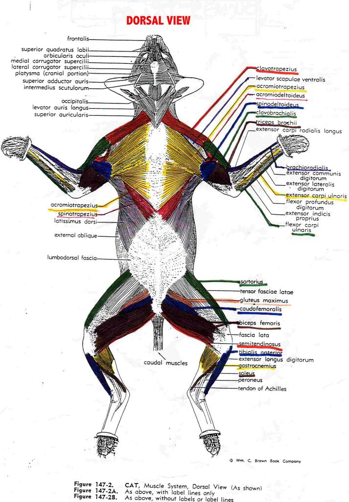
E. Muscles of the Back and Shoulder 1.Refer to Figure C1.4a to locate the following superfi- cial muscles: •Trapezius muscles—The cat has three separate mus- cles,compared with a single human trapezius. —Clavotrapezius —Acromiotrapezius —Spinotrapezius •Deltoid muscles—The cat has three separate deltoid muscles,compared with one in humans.
PNB communique: Cat muscles lab info + Quiz 3 guide
Muscles of the cat : simplebooklet.com

Pin on Animal ref
Muscles are how we move and live. All movement in the body is controlled by muscles. Some muscles especially 5 Cat Muscle Anatomy Diagram work without us thinking, like our heart beating, while other muscles are controlled by our thoughts and allow us to do stuff and move around. There are over 650 muscles in the human body.
Blue-eyed cat portrait
07/09/2021 · Dog front leg muscles anatomy You will find the extrinsic and intrinsic muscles in the front leg of a dog. I will show you the essential muscles from the dog front leg anatomy with a labeled diagram. Fine, if you want to know more about the dog muscle anatomy, you may read the article here.

Muscles of the cat : simplebooklet.com
Chapter 8: Muscular System. This chapter is divided into three main sections: muscle basics and cellular components, naming of the muscles, and cat muscles with dissection. Be prepared to spend a fair amount of time on this unit.

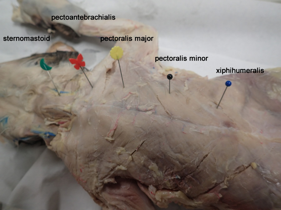
Pectoralis Muscles of the Cat
Cat Muscles of the Back (color) Image shows the dorsal (back) side of a cat with well defined muscles. Color the muscles to help learn them. Muscles include the trapezius, latissimus dorsi, gluteus maximus, triceps, biceps, gastrocnemius, and sartorius. T Tabitha Kohring nursing school

Cat Muscles
Cat Anatomy Dissection Guide Superficial Muscles Ventral View pectoantebrachialis Dorsal View clavotrapezius pectoralis major acromiotrapezius pectoralis minor spinotrapezius xiphihumeralis latissiumus dorsi external oblique clavobrachialis internal oblique acromiodeltoid transversus abdominis spinodeltoid ...

Munchkin Cat Muscle Anatomy by TheDragonofDoom on DeviantArt
Cat References Day 1 Terminology & External/Internal Anatomy. Pictures from iBook; Cat Skeleton Tutorial-Kenyon College; Day 2&3 Muscular System. Muscle Tables; Cutaneous, Shoulder & Back Muscles

Cat Muscles Labeled | Anatomy and Physiology Background ...
Human Muscles Labeled Diagram For Kids Infographics Muscle. C Ous Atae M Figure 311 Illustrates The Ven Cheggcom. Royalty Free Stock Illustration Of Diagram Dorsal Ventral Cavities. A Motor And A Brake Two Leg Extensor Muscles Acting At The Same. Muscle Diagrams To Label Diagram Dorsal Cat Oasissolutionsco.

cat anatomy muscles diagram | Vet School | Pinterest | Cat ...
Find the rectus femoris by separating the sartorius and biceps femoris. The rectus femoris is a deep muscle between the two. Locate the palmaris longus and the brachioradialis in the upper limb of the cat.

1000+ images about cat muscles on Pinterest | Cats, The ...

CatSkeleton.png (1163×741)

Cat Muscles

Pin di muscles

Cat Muscles

Cat Anatomy - or Catnatomy! A Look Inside Your Cat

22 best images about Anatomy..Creating a New Aspect. on ...

Black Rabbit, Byker Farm, Ouseburn Valley, Newcastle Upon Tyne, Tyne & Wear, England.

Cat muscles | Vet tech | Pinterest

Cat Muscles

Muscle Diagram Quiz — UNTPIKAPPS
Cat Muscles

Human Anatomy & Physiology 1

Cat Muscles Lab Guide
We adopted our white cat, Neige, in September 2017, when she was only two months old. One day, while setting up our studio, she decided to go right in the middle of it, stood up, and looked right at us, as if she wanted someone to take her photo. So, we did. The photo was simply taken with a smartphone, as I’m sure she wouldn’t have waited for us to set up our gear to take a photo of her. The lighting and the pose were already perfect, hence I didn’t hesitate to take that photo and capture the moment.

Msucles Cat Dissection Diagrams in 2020 | Muscle diagram ...

cat muscles | Dorsal View of Superficial Muscles of the ...
0 Response to "39 cat muscles labeled diagram"
Post a Comment