37 drag the labels onto the diagram of the cns meninges.
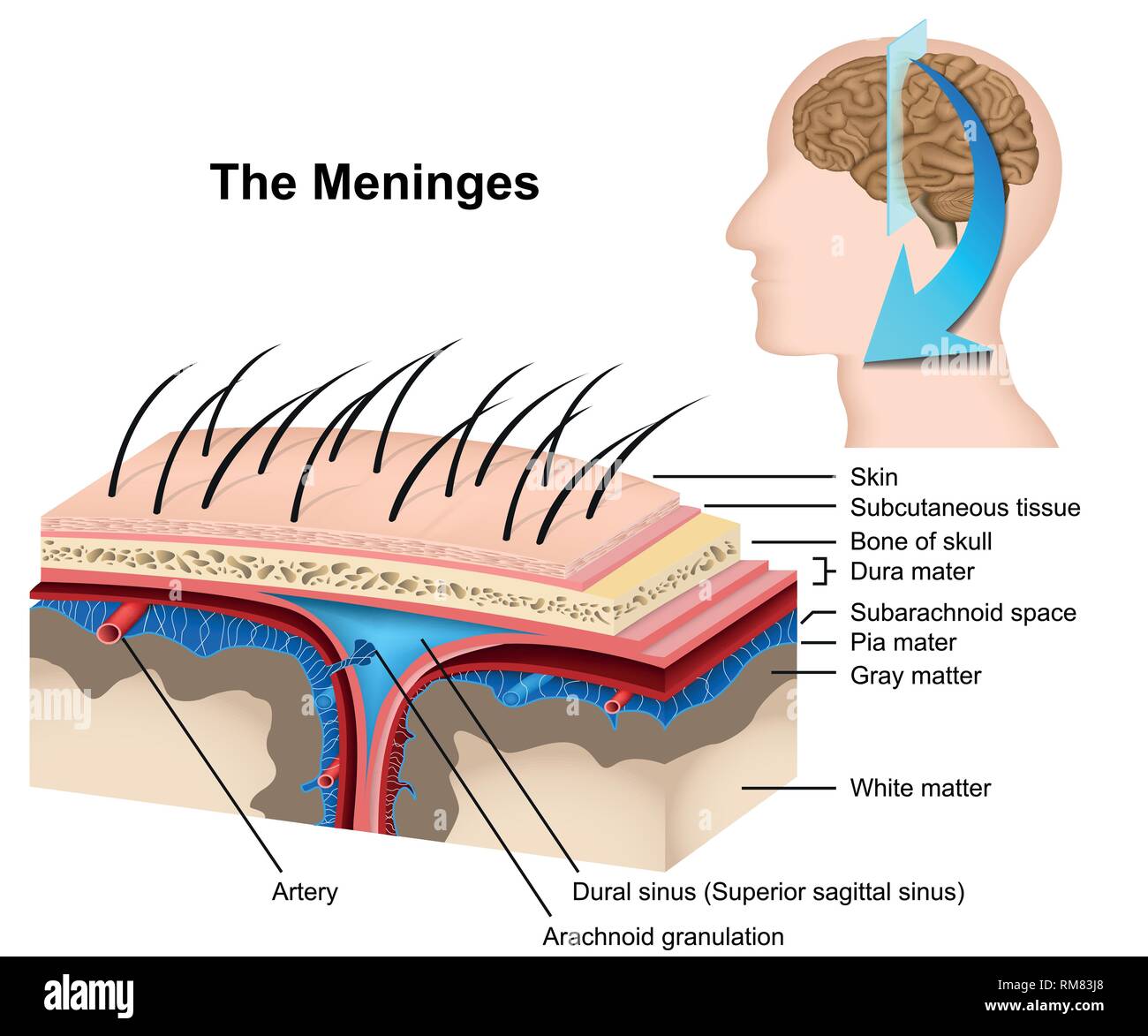
Meninges that surround the CNS consist of an outer fibrous sheet of dura mater (pachymeninx) that is also the inner periosteum of the skull. As the foramina of Magendi and Luschka develop, one continuous CSF system evolves. Due to the lack of arachnoid granulations during foetal life, it is most... Jul 01, 2019 · The meninges functions primarily to protect and support the central nervous system (CNS). It connects the brain and spinal cord to the skull and spinal canal. The meninges forms a protective barrier that safeguards the sensitive organs of the CNS against trauma. It also contains an ample supply of blood vessels that deliver blood to CNS tissue.
Drag the arrows onto the diagram below to indicate the direction that DNA polymerase III moves along the parental (template) DNA strands at each of the two replication forks. 5 hours ago Art-labeling Activity: Brain, Cranium, and Meninges (Close-up View of Cranial Meninges) Part A Drag the labels...

Drag the labels onto the diagram of the cns meninges.
We review their content and use your feedback to keep the quality high. 100% (11 ratings) Answer The label is indicated from RIGHT SIDE of image to …. View the full answer. Transcribed image text: Part A Drag the labels onto the diagram to identify the spinal nerve roots and meninges Reset Help Ventral Pia mater Meninges Dorsal root Dura mater. Development of the Central Nervous System (CNS) includes development of the brain, spinal cord, optic and auditory systems, as well as surrounding supporting cells including ependymal cells, astrocytes, oligodendrocytes and microglia. Label the meninges of the brain and spinal cord.Part ADrag the labels onto the diagram of the CNS meninges.Central canalAqueduct of SylviusArachnoid villusCranial vein valves. 4/11/2017Chapter 910/10CorrectScore Summary:Your score on this assignment is 97.1%.You received...
Drag the labels onto the diagram of the cns meninges.. Drag and drop the descriptive labels of events into the correct sequence at the chemical synapse. 1 4 The Somatic Nervous System Neuroscience Canadian 1st Edition. Label the parts of the neuromuscular junction. Cns central nervous system 7. Drag the labels onto the diagram to... The central nervous system (CNS) consists of the brain and spinal cord. This body system is responsible for integrating and coordinating the activities of the entire body. Overview. The central nervous system can be thought of as the coordination and integration system within organisms. Reset Help central cand matrix Group 2 lacuna Group 2 Group 2 osteocyte in lacuna Group 2 C chondrocyto Group 2 bono (osseous tissue) Group 1 Bone tissue differentiates from other tissues because of the mineralized extracellular matrix. This tissue provides support and protection due to the... After each piece of the lagging stand is complete it is released from dna polymerase. Na is entering the cell. 3 3 Eukaryot...
Drag the labels onto the diagram to identify the cranial meninges and associated structures. Drag the labels to identify the landmarks and features on one of the cerebral hemispheres. Drag the labels onto the diagram to identify the origins of the cranial nerves (I - VI). Each muscle spindle is a feedback mechanism that detects muscle length and any changes to muscle length by increasing the number of electri... The spinal cord and meninges are contained in the spinal channel, ... Drag the labels onto the diagram to identify the components of the somatic nervous ...1 answer · 0 votes: Spinal cord is the part of central nervous system along with brain. The spinal cord is a long, delicate tubelike structure that starts toward the finish ... The meninges are the fibrous covering of the central nervous system (CNS) which contain vastly heterogeneous cell types within its three layers (dura While the meninges were historically seen as a purely structural support for the CNS and protection from trauma, the emerging view of the meninges...
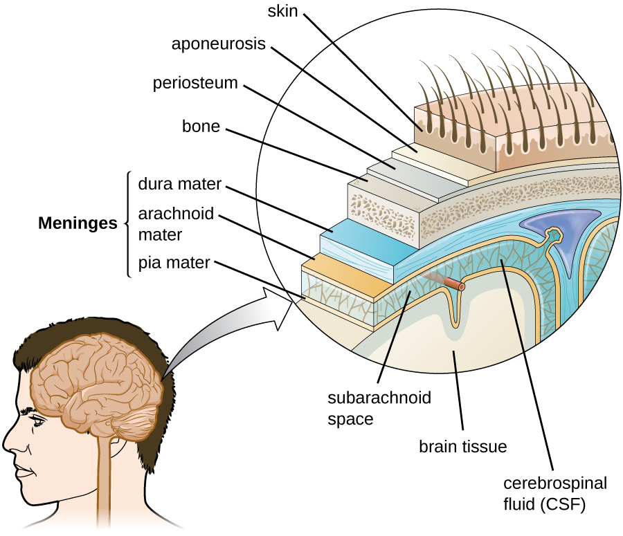
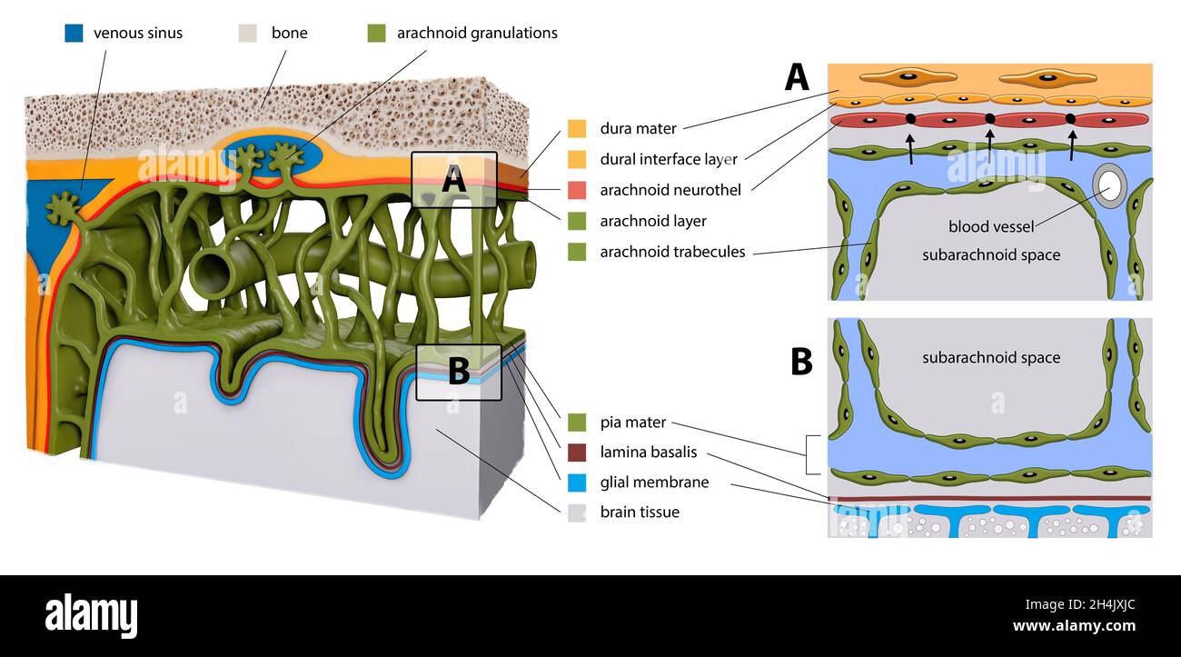
Meninges. The brain and the spinal cord Spinal cord The spinal cord is the major conduction pathway connecting the brain to the body; it is part of the CNS. of the meninges, and the etiology can be elicited by examining CSF, which is contained within the subarachnoid space. meninges, three membranous envelopes—pia mater, arachnoid, and dura mater—that surround the brain and spinal cord. The pia mater is the meningeal envelope that firmly adheres to the surface of the brain and spinal cord. It is a very thin membrane composed of fibrous tissue covered on its outer... In the perivascular space and subdural meninges, A single-cell atlas of mouse brain macrophages. Recent work has revealed that fibroblasts play crucial roles in fibrotic scar formation in the CNS Here, we are going to focus on the role of the peripheral immune system and its crosstalk with CNS in the... The meninges refer to the membranous coverings of the brain and spinal cord, comprised of the dura, arachnoid and pia mater. Provide a supportive framework for the cerebral and cranial vasculature. Acting with cerebrospinal fluid to protect the CNS from mechanical damage.
14.2 Blood Flow the meninges and Cerebrospinal Fluid ...
Cns central nervous system 7. Can you name the label the parts of the neuromuscular junction. Drag and drop the descriptive labels of events into the correct sequence at the chemical synapse.

Pacific Medical Training - Nervous System
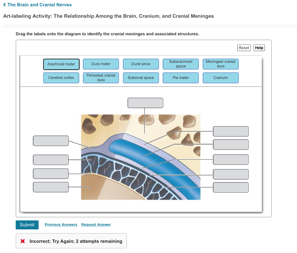
Drag the labels onto the diagram to identify the cranial meninges and associated structures. 1. dura mater 2. subarachnoid space 3. pia mater 4. cerebral cortex 5. cranium 6. periosteal cranial dura 7. dural sinus 8. meningeal cranial dura 9. subdural space 10. arachnoid mater.

lab 7 (exercise 14) Flashcards | Quizlet
The central nervous system (CNS) consists of the brain, spinal cord, spinal fluid and neurons, which transmit signals from the brain to other body Efferent neurons are mainly located in the peripheral nervous system, but their cell bodies orginate in the CNS. Many incoming signals from the CNS...
Anatomy and Physiology Lab I†on OpenALG
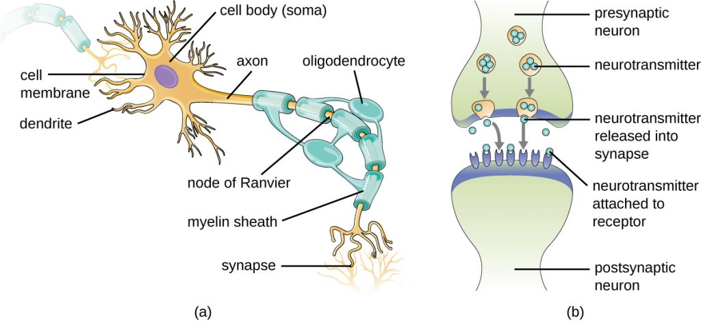
The central nervous system (CNS) consists of the brain and spinal cord. It connects to the peripheral nervous system (PNS), a network of nerves that Figure 2. The layers of tissue surrounding the human brain include three meningeal membranes: the dura mater, arachnoid mater, and pia mater...
SAY: Welcome to Module 1: Anatomy & Physiology of the Brain ...
The central nervous system (CNS) is the part of the nervous system consisting primarily of the brain and spinal cord. The CNS is so named because the brain integrates the received information and coordinates and influences the activity of all parts of the bodies of bilaterally symmetric animals—that...
27.1 Anatomy of the Nervous System – Microbiology: Canadian ...
2- Histology of spinal cord-CNS-Second year-New edition 2021. Neurology | Anatomy & Function of the Cerebellum.

lab 7 (exercise 14) Flashcards | Quizlet
The meninges (singular meninx; Greek, meninx = membrane) are a complex connective tissue surrounding the central nervous system (brain and spinal cord). The 3 layers from the central nervous outward are: pia mater, arachnoid mater, and the dura mater.
Duke Neurosciences - Lab 1: Surface Anatomy of the Brain
pia mater onto the brain ct covering arachnoid mater tough ct double layer outer periosteal, inner meningeal connective tissue coverings of. External cover that protects brain & spinal cord Ń Extensions into cranial cavity limit movement of the brain `.
11.6 Nervous System – Concepts of Biology – 1st Canadian Edition
Anatomy and Physiology. Anatomy and Physiology questions and answers. Part A Drag the labels onto the diagram to identify the cranial meninges and associated structures. Reset Hel Subdural space Pia mater Subarachnoid space Dural sinus Cranium Dura mater (meningeal layer) Dura mater (periosteal layer) Arachnoid mater Cerebral cortex QUID Submit Request Answer.

Spinal Meninges | Draw it to Know it
The Central Nervous System (CNS) consists of: The spinal cord Integrates and processes information Can function with the brain Can function independently. Chapter 14 - The Nervous System: The Spinal Cord and Spinal Nerves $100 $200 $300 $400 $500 $100$100$100 $200 $300...

Topic 11 Divisions of the Nervous System - ppt download
Meninges-brain interface: signals from the meninges regulate development of the CNS 1041. Infiltration of the meninges is such a common complication of acute leukaemia that routine treatment of acute leukaemia in childhood includes regular prophylactic measures to prevent its occurrence.

Lab Activity Chapter 20.pdf - Lab Activity Chapter 20 Lab ...
Drag and drop the text labels onto the boxes next to the heart diagram. 1 pulmonary ventilation the total exchange of air usually measured in litersminute and 2 alveolar ventilation the effective ventilation of the alveoli in which gas exchange with the blood actually takes place.

Sheep brain dissection | Human Anatomy and Physiology Lab ...
Meningeal blood supply. This is a branch of the maxillary artery , which, despite its name, primarily supplies the calvarium rather than the meninges 1,2 . Clinical importance is given to this artery due to its location in the extradural space and the proximity of its anterior division to the pterion , making it...

A&P Chapter 11 Nervous System 2 Homework Flashcards | Quizlet
The central nervous system is the supreme command center of the human body. Learn about its anatomy and function now at Kenhub! The CNS consists of two organs which are continuous with each other; the brain and spinal cord. They are enveloped and protected by three layers of meninges...

Chapter 12 HW The Central Nervous system Flashcards | Quizlet
Label the meninges of the brain and spinal cord.Part ADrag the labels onto the diagram of the CNS meninges.Central canalAqueduct of SylviusArachnoid villusCranial vein valves. 4/11/2017Chapter 910/10CorrectScore Summary:Your score on this assignment is 97.1%.You received...

9 BRAIN ideas | brain, dura mater, brain anatomy
Development of the Central Nervous System (CNS) includes development of the brain, spinal cord, optic and auditory systems, as well as surrounding supporting cells including ependymal cells, astrocytes, oligodendrocytes and microglia.

A&P2 Lab 2 HW Flashcards | Quizlet
We review their content and use your feedback to keep the quality high. 100% (11 ratings) Answer The label is indicated from RIGHT SIDE of image to …. View the full answer. Transcribed image text: Part A Drag the labels onto the diagram to identify the spinal nerve roots and meninges Reset Help Ventral Pia mater Meninges Dorsal root Dura mater.

A&P2 Lab 2 HW Flashcards | Quizlet

A&P2 Lab 2 HW Flashcards | Quizlet
The Nervous System
Meninges Illustration High Resolution Stock Photography and ...
The Nervous System Introduction The Meninges The Meninges The ...
Anatomy of the Head and Neck | SpringerLink

Mastering A&P Chapter 13 Flashcards | Quizlet

lab 7 (exercise 14) Flashcards | Quizlet

lab 7 (exercise 14) Flashcards | Quizlet

Anatomy 2220 Brain and SP Lab Review Flashcards | Quizlet

Meninges & CSF - Neuroanatomy Flashcards | Draw it to Know it
Chapter 14 Lecture Outline

lab 7 (exercise 14) Flashcards | Quizlet
Anatomy Exam 2 Flashcards - Easy Notecards
27.1 Anatomy of the Nervous System – Microbiology: Canadian ...

Meninges & CSF - Histology | Draw it to Know it

Drag the appropriate labels to their respective targets ...
Solved K The Brain and Cranial Nerves Art-labeling Activity ...
Arachnoid Mater High Resolution Stock Photography and Images ...
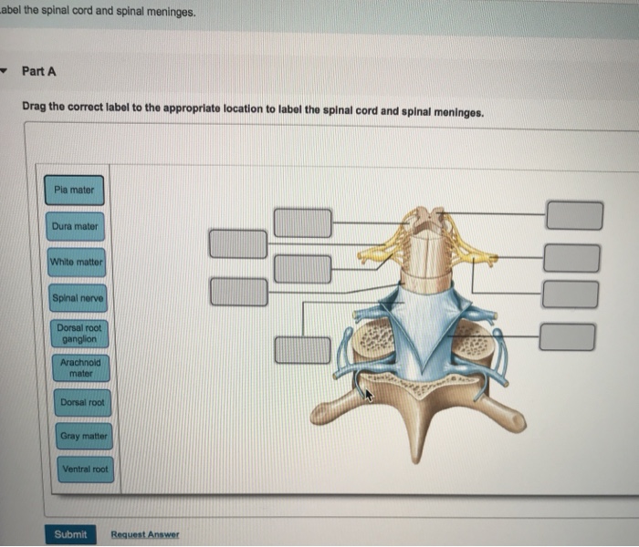
Solved abel the spinal cord and spinal meninges. Part Drag ...
0 Response to "37 drag the labels onto the diagram of the cns meninges."
Post a Comment