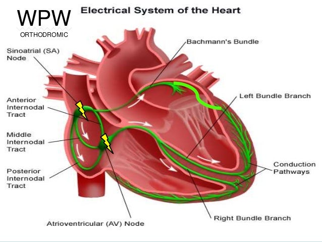
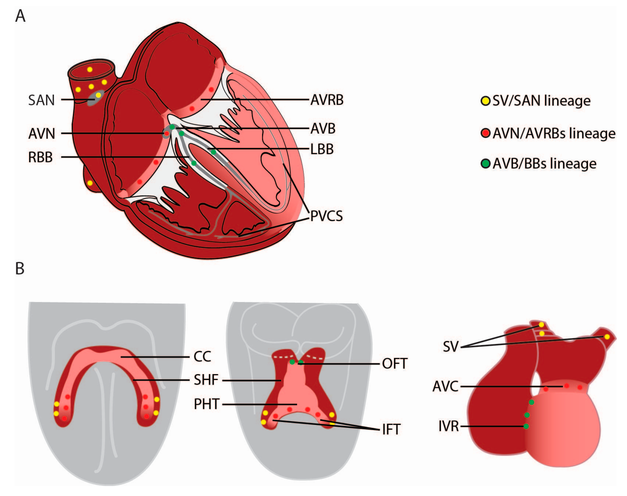
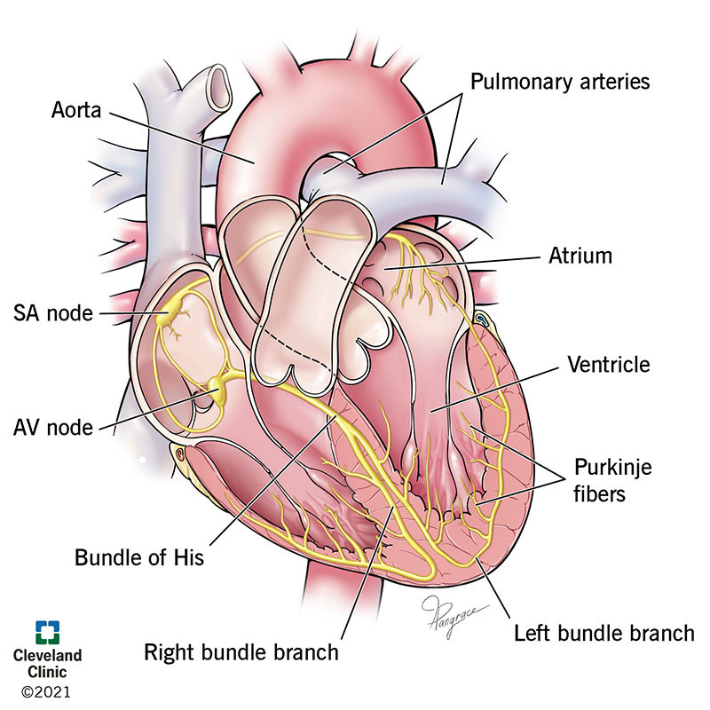
35 cardiac conduction system diagram
Cardiac Conduction System · The sino-atrial (SA) node · The atrio-ventricular (AV) node · The bundle of His · The left and right bundle branches · The Purkinje ... Cardiac diastole is the period of the cardiac cycle when, after contraction, the heart relaxes and expands while refilling with blood returning from the circulatory system. Both atrioventricular (AV) valves open to facilitate the 'unpressurized' flow of blood directly through the atria into both ventricles, where it is collected for the next contraction.
Lower respiratory tract organs. Trachea: Also known as the windpipe this is the tube that carries air from the throat into the lungs. It ranges from 20-25mm in diameter and 10-16cm in length. The inner membrane of the trachea is covered in tiny hairs called cilia, which catch particles of dust which we can then remove through coughing.

Cardiac conduction system diagram
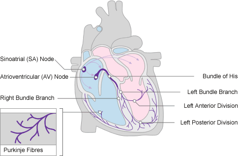
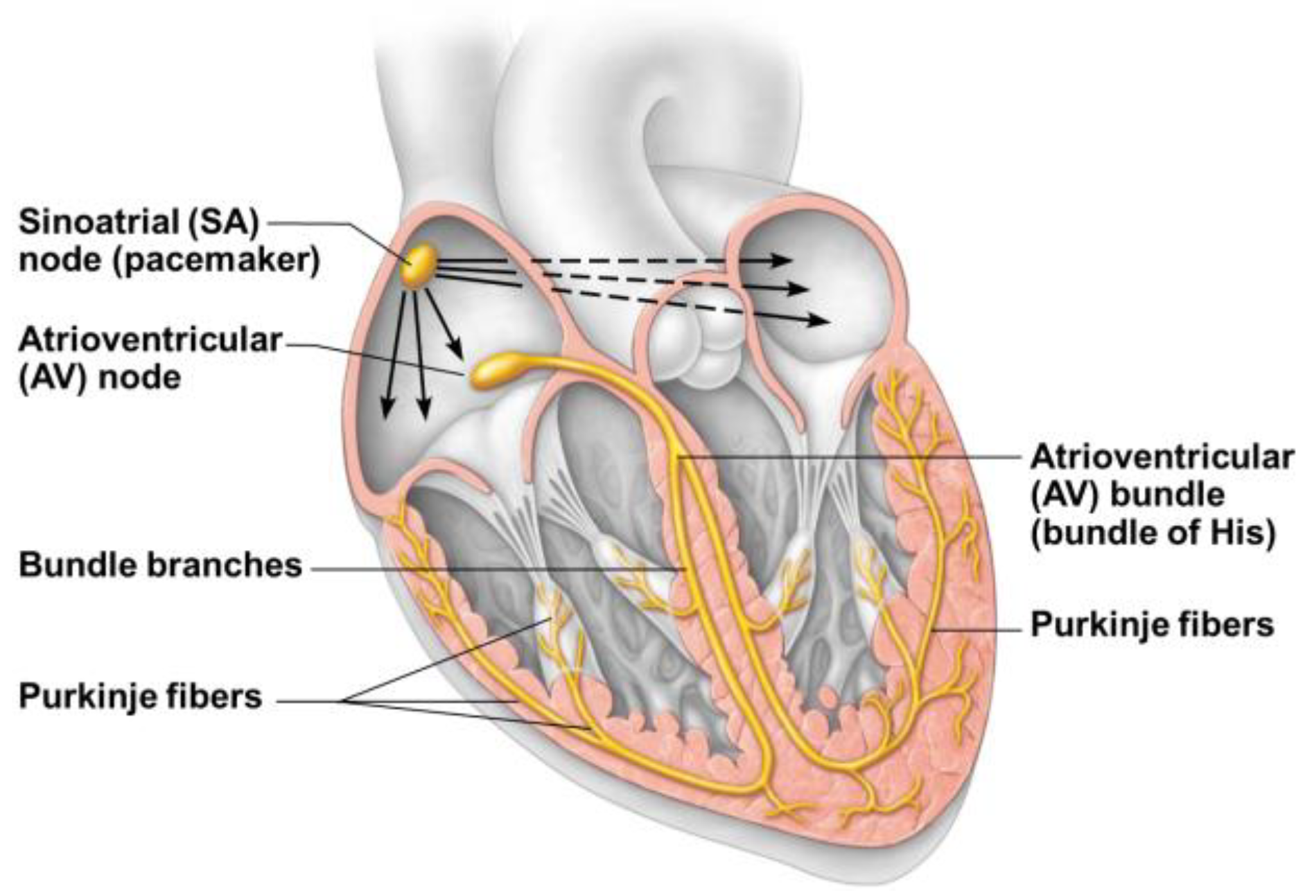
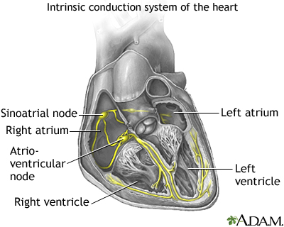
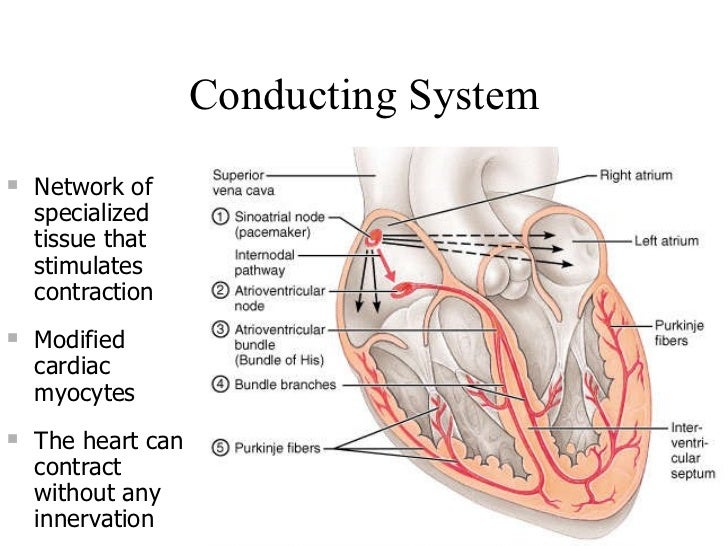
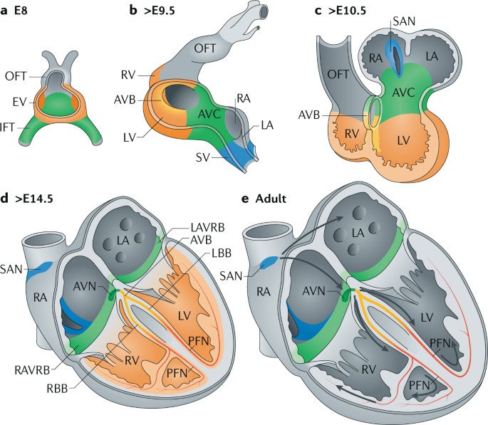
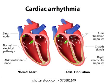
A fully developed adult heart maintains the capability of generating its own electrical impulse, triggered by the fastest cells, as part of the cardiac conduction system. The components of the cardiac conduction system include the sinoatrial node, the atrioventricular node, the atrioventricular bundle, the atrioventricular bundle branches, and the Purkinje cells. Your heart (cardiac) conduction system sends the signal to start a heartbeat. It also sends signals that tell different parts of your heart to relax and contract (squeeze). This process of contracting and relaxing controls blood flow through your heart and to the rest of your body. The electrical conduction system of the heart transmits signals generated usually by the sinoatrial node to cause contraction of the heart muscle.The pacemaking signal generated in the sinoatrial node travels through the right atrium to the atrioventricular node, along the Bundle of His and through bundle branches to cause contraction of the heart muscle.
Cardiac conduction system diagram. 7 Jul 2020 — A network of specialized muscle cells is found in the heart's walls. These muscle cells send signals to the rest of the heart muscle causing a ... 28 Jun 2016 — The conducting system of the heart consists of cardiac muscle cells and conducting fibers (not nervous tissue) that are specialized for ... The cardiac conduction system is a collection of nodes and specialised conduction cells that initiate and co-ordinate contraction of the heart muscle. 28-10-2021 · The cardiac cycle is defined as a sequence of alternating contraction and relaxation of the atria and ventricles in order to pump blood throughout the body. It starts at the beginning of one heartbeat and ends at the beginning of another. The process begins as early as the 4th gestational week when the heart first begins contracting.. Each cardiac cycle has a diastolic phase (also called ...
by VM Christoffels · 2009 · Cited by 176 — The conduction system of the heart initiates and coordinates the electric signal that causes the rhythmic and synchronized contractions of ... 06-02-2021 · Fig 2 – Diagram showing the action potential in cardiac pacemaker cells and the main ion movements at each stage. Control by the Autonomic Nervous System The autonomic nervous system (ANS) alters the slope of the pacemaker potential, in order to alter heart rate. The heart's pumping action is regulated by an electrical conduction system that coordinates the contraction of the various chambers of the heart. Diagram of ... 27-09-2020 · Outline of the Cardiac Conduction System (without the heart muscle). The sinoatrial (SA) node is the normal site of origin of the electrical impulse (action potential) that stimulates heart muscle to contract. The SA node is located in the upper region of the right atrium.
05-10-2020 · Easily learn the conduction system of the heart using this step-by-step labeled diagram. The cardiac conduction system is the electrical pathway of the heart that includes, in order, the SA node, AV node, bundle of His, bundle branches, and Purkinje fibers. … 21-02-2021 · At rest the heart pumps around 5l of blood around the body every minute, but this can increase massively during exercise. In order to achieve this high output efficiently the heart works through a carefully controlled sequence with every heart beat – … The electrical conduction system of the heart transmits signals generated usually by the sinoatrial node to cause contraction of the heart muscle.The pacemaking signal generated in the sinoatrial node travels through the right atrium to the atrioventricular node, along the Bundle of His and through bundle branches to cause contraction of the heart muscle. Your heart (cardiac) conduction system sends the signal to start a heartbeat. It also sends signals that tell different parts of your heart to relax and contract (squeeze). This process of contracting and relaxing controls blood flow through your heart and to the rest of your body.
A fully developed adult heart maintains the capability of generating its own electrical impulse, triggered by the fastest cells, as part of the cardiac conduction system. The components of the cardiac conduction system include the sinoatrial node, the atrioventricular node, the atrioventricular bundle, the atrioventricular bundle branches, and the Purkinje cells.
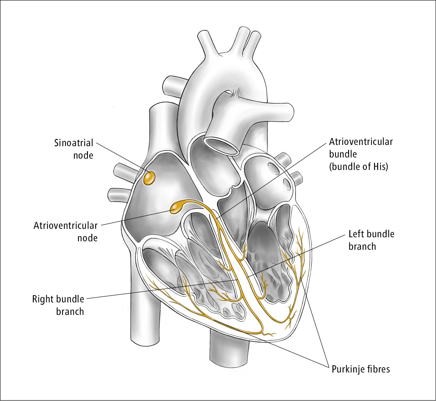
Figure 031 6254 Cardiac Conduction System Illustration Courtesy Of Dr Shannon Zhang Mcmaster Textbook Of Internal Medicine

Electrical Conduction System Of The Heart Saltatory Conduction Anatomy Bundle Of His Heart Angle Text Hand Png Pngwing









0 Response to "35 cardiac conduction system diagram"
Post a Comment